Endoscope used for prostate operations

An endoscope, prostate technology, applied to endoscope, surgery, surgical forceps and other directions, can solve the problems of difficult operation of electric cutting ring, adverse effects on systemic hemodynamics, increased workload, etc., to achieve the effect of convenient surgery

Inactive Publication Date: 2010-07-28
SHANGHAI KINDBEST MEDICAL TECH
View PDF3 Cites 12 Cited by
  • Summary
  • Abstract
  • Description
  • Claims
  • Application Information

AI Technical Summary

Problems solved by technology

[0003] However, this "gold standard", such as the current resectoscope surgery, has the following disadvantages: 1. It requires a large amount of expensive lavage fluid, and a prostate resection operation requires about 30,000 to 40,000 ml; 2. Intraoperative bleeding , easy to dissolve with lavage fluid, resulting in blurred vision, frequent water release and irrigation, which not only wastes a lot of water quality, but also increases the pressure in the bladder due to a large amount of perfusion fluid during the operation, which is prone to water intoxication and endangers life; 3. Liquid perfusion cannot be controlled The pressure in the bladder frequently makes the bladder high and low, which has adverse effects on the hemodynamics of the whole body; 4. The water used in the operation often contains viruses and bacteria, which will cause environmental pollution if it overflows into the operating room or flows into the sewer, and even causes medical staff Pollution
[0004] However, the existing prostate surgery often requires the use of electric cutting rings, and the application of electric cutting rings has the following problems: 1. For resection of benign prostatic hyperplasia, electric cutting rings need to be cut one by one, and a blood vessel needs to be cut multiple times before hemostasis; 2. Ring cutting requires the operation of digging out, and t

Method used


Image

  • Endoscope used for prostate operations
  • Endoscope used for prostate operations
  • Endoscope used for prostate operations

Examples


Embodiment Construction

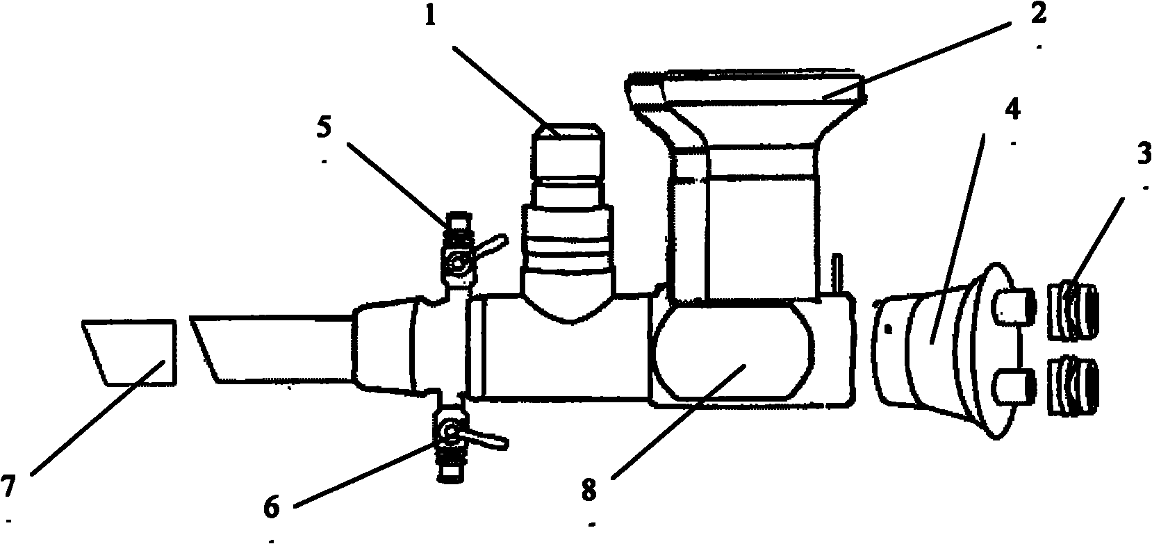
[0017] figure 1 It is a schematic diagram of the structure of the surgical endoscope of the present invention. The following embodiments take intra-urethral bladder surgery as an example, and further illustrate the present invention with reference to the accompanying drawings.

[0018] An endoscope for prostate surgery, comprising: an endoscope body 8, on which a light source interface 1 and an observation mirror 2 are mounted, and is characterized in that: the endoscope body 8 and the observation mirror 2 are perpendicular to each other, the back of the endoscope body 8 is sequentially connected with a closer 4 and an anti-reflux cap 3, and the front of the endoscope body 8 is provided with a treatment channel 7 and carbon dioxide for the passage of surgical instruments The gas inlet valve 6 can place multiple replaceable instruments in the cavity of the endoscope body 8.

[0019] The replaceable instruments placed in the cavity of the endoscope body 8 are rugby-ball-shaped peelin...


PUM


Abstract

The invention discloses an endoscope used for prostate operations, which comprises an endoscope body, wherein a light source port and an observation mirror are installed on the endoscope body. The endoscope is characterized in that the endoscope body is perpendicular to the observation mirror; a closer and an anti-reflux sealing cap are sequentially connected at the rear part of the endoscope body; a cavity on which changeable surgical instruments are put is arranged in the endoscope body; a treatment channel for the surgical instruments to pass through is connected at the front part of the endoscope body; and two carbon dioxide inlet valves are arranged at the front part of the endoscope body. The closer is provided with more than three pore canals for the surgical instruments to pass through for operation. The endoscope can be used in prostatectomy, resection of bladder tumors, vesical diverticulectomy, ureterocystostom and the like, and has the good effects that the endoscope can provide the doctor with a clear picture of the affected part before the operation, and with the endoscope, the doctor can operate with both hands to finish the highly difficult operations of resection, suture and the like, thus providing convenience for the doctor to conduct medical operations.

Description

Technical field [0001] The invention relates to a minimally invasive surgical medical equipment, in particular to an endoscope for prostate surgery. Background technique [0002] In the past 70 years, minimally invasive techniques of transurethral resection using transurethral resection have been used for bladder tumor diseases and prostate hyperplasia diseases. Called transurethral resection microscope. The scope uses a circular electrocutter under peeping to cut tissue one by one, and has electrocoagulation effect. Observation of the visual field is achieved by infusion of 5% mannitol or 5% glucose. In the past two decades, more than 95% of hospitals at home and abroad have adopted this method as the "gold standard" for the treatment of benign prostatic hyperplasia, and have basically abandoned open surgery for the treatment of benign prostatic hyperplasia. [0003] However, this "gold standard", for example, the current resection surgery has the following shortcomings: 1. A l...

Claims


Application Information

Patent Timeline
IPC IPC(8): A61B17/94A61B18/14
CPCA61B2018/00577A61B1/018A61B1/307A61B1/015A61B2018/00547A61B2018/048
Inventor 林超段孝敏
Owner SHANGHAI KINDBEST MEDICAL TECH
Why Eureka
Try Eureka