Optical biosensor for detecting brain natriuretic peptide (BNP) and preparation method of reagent thereof

A biosensor and brain natriuretic peptide technology, applied in the field of biosensing, can solve the problems of the preparation method of the brain natriuretic peptide optical biosensing technology, which affects the detection speed and sensitivity, and achieve simple and fast detection, easy promotion and operation convenient effect

Inactive Publication Date: 2010-08-04
INST OF ELECTRONICS CHINESE ACAD OF SCI
View PDF2 Cites 15 Cited by
  • Summary
  • Abstract
  • Description
  • Claims
  • Application Information

AI Technical Summary

Problems solved by technology

Research shows that reducing detection cost will affect detection speed and sensitivity
At present, domestic research on BNP is mainly based on the clin

Method used


Image

  • Optical biosensor for detecting brain natriuretic peptide (BNP) and preparation method of reagent thereof
  • Optical biosensor for detecting brain natriuretic peptide (BNP) and preparation method of reagent thereof
  • Optical biosensor for detecting brain natriuretic peptide (BNP) and preparation method of reagent thereof

Examples


Embodiment 1

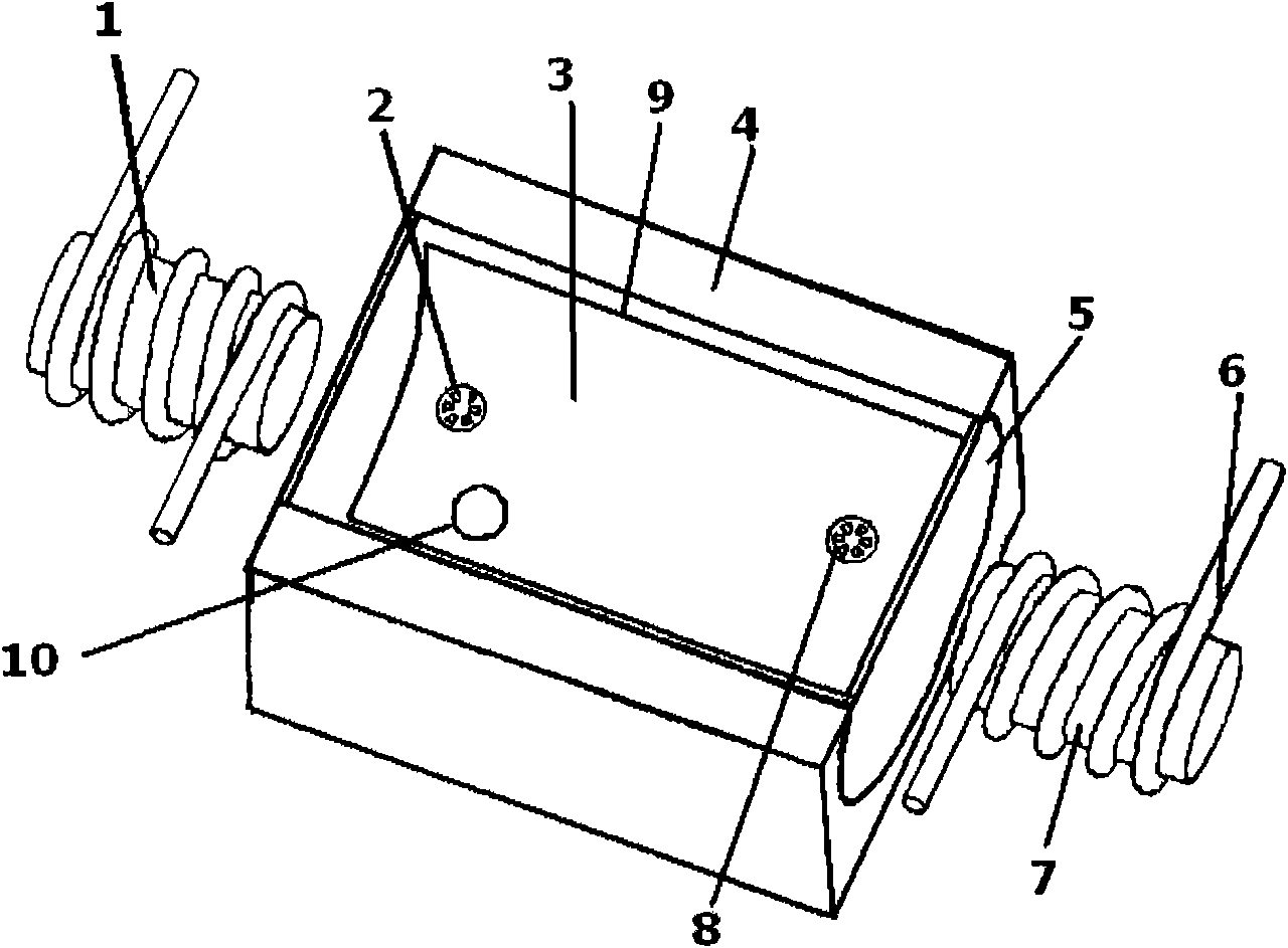
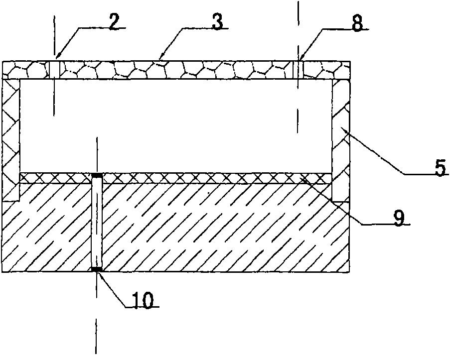
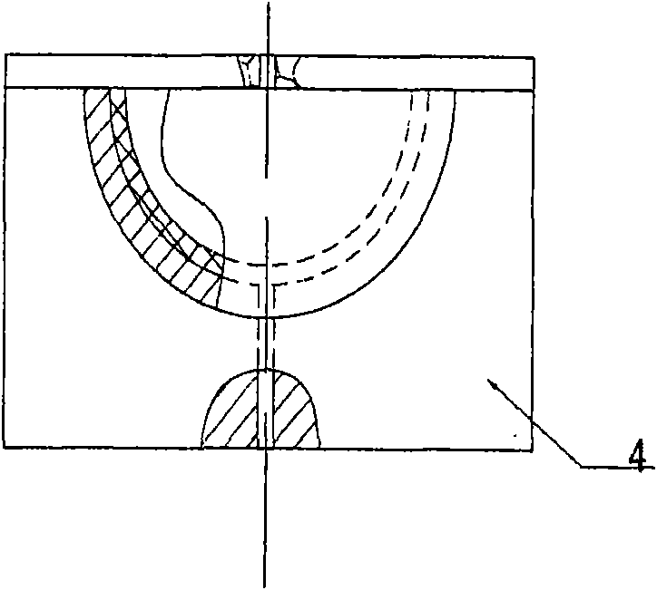
[0042] figure 1 2(a), (b), and (c) are schematic diagrams of the magnetic nanoprobe-based BNP optical biosensor designed by the present invention. First, a micro-channel with a length of 20 mm, a width of 5 mm, and a depth of 4 mm is etched on a silicon base using a micro-electro-mechanical system (MEMS) process, and the top of the channel is bonded with a glass with excellent light transmission ( A cover sheet) 3, a sample injection hole 2 and a sample injection hole 8 are arranged on the glass (cover sheet) 3, and a sample outlet 10 communicating with the microchannel is arranged at the bottom of the silicon base 4. Assemble a polymer carboxymethylcellulose (CMC) nanomaterial layer 9 on the inner surface of the channel in the reaction zone, place it in a drying oven at 37° C. for 30 minutes, and take it out for use.

[0043] Add reaction reagent then, drip 50 μ l brain natriuretic peptide magnetic nanoprobe solution from injection hole 2, drip 20 μ l brain natriuretic pepti...

Embodiment 2

[0045] 1) Preparation of BNP-specific probes

[0046] Take 200 μl of amino-terminal magnetic nanoparticles and place them in a 2 ml centrifuge tube, add 600 μl of 0.03 mol / L pyridine solution, and shake to mix. Remove the supernatant by magnetic separation with a magnetic stand, and repeat the above operation twice. Then, 200 μl of 10% glutaraldehyde was added to the centrifuge tube, and placed in a constant temperature shaker at 37° C. at 220 rpm for 30 minutes to shake and react for uniform mixing. Magnetic separation was performed to remove unreacted glutaraldehyde, and finally washed three times with 200 μl Tris-HCl buffer solution (0.05 mol / L; pH=7.10-9.00).

[0047] Dissolve the BNP antibody in PBS buffer, and prepare an antibody solution with a concentration of 0.22-2.0 mg / mL. Take 400 μl of the BNP antibody solution and add it to 200 μl of the above-treated magnetic nanoparticles, and shake and react at 180 rpm in a constant temperature shaker at 37° C. for 6 hours. ...

Embodiment 3

[0052] 1) Preparation of BNP-specific probes

[0053] Such as image 3 , use a pipette to pipette 200μl (equivalent to 1mg) of magnetic nanoparticles labeled with avidin on the surface, place them in a 2ml centrifuge tube, add 1000μl Tris-HCl buffer (containing 1mmol / LEDTA, 1mol / L NaCl, pH =7.4), shake gently to mix the magnetic particles, magnetically separate, and wash twice. The above-mentioned magnetic particles were dissolved with 100 μl of PBS buffer.

[0054] 100 μl of biotin-labeled BNP antibody was dissolved in 800 μl of Tris-HCl buffer, and the antibody concentration was 0.1875 mg / mL. The above antibody solution was added to the activated magnetic particles, placed in a shaker at 37°C and 150 rpm for 30 min, and subjected to magnetic separation, and the supernatant was removed. Add 1 ml of PBS buffer solution (containing 0.05% Tween-20 and 0.1% BSA) into the centrifuge tube after the coupling reaction, shake gently to mix, magnetically separate, and operate twice....


PUM

PropertyMeasurementUnit
Depthaaaaaaaaaa
Diameteraaaaaaaaaa

Abstract

The invention discloses an optical biosensor for detecting brain natriuretic peptide (BNP) and a preparation method of a reagent thereof, relating to the biosensing technology. A sensor reaction zone is a capillary channel filled with magnetic nano probe solution, the inner surface of the channel is assembled with a layer of hydrophilic nano material, the upper surface of the channel is an euphotic insulating encapsulation layer with an injection hole, the side surface of the channel is encapsulated by plastics, the two ends thereof encapsulated with plastics are respectively provided with a controllable electromagnet. The preparation method of the reagent is as follows: preparing magnetic nano probes, fully reacting the magnetic nano probes, a brain natriuretic peptide (BNP) sample to bedetected and specificity BNP antigen marked by a marker under the united action of the electromagnet and the capillary channel, forming a compound of magnetic nano probes, BNP antigen to be detected and marked antibody, and adding substrate for catalytic luminescence or exciting luminescent quantum dots for luminescence. By the sensor detection system, the BNP concentration can be rapidly detected. The sensor in the invention can rapidly detect the BNP concentration in a sample, and has the characteristics of high specificity, high sensitivity, a great quantity of saved biological reagent andthe like.

Description

technical field [0001] The invention relates to the technical field of biosensing, and relates to an optical biosensor for rapidly detecting the concentration of a heart failure marker brain natriuretic peptide trace protein and a preparation method for the reagent used. The sensor can quickly detect the concentration of total brain natriuretic peptide. Background technique [0002] Brain natriuretic peptide is a hormone-like short peptide secreted by the ventricle. The active peptide is brain natriuretic peptide-32. The ring structure, which is the functional domain of BNP. Brain natriuretic peptide binds to its corresponding receptor through this conformation and plays a role in many pathophysiological processes. The number of brain natriuretic peptides is very small, and the more serious the heart failure is, the higher the value of brain natriuretic peptides is. Therefore, the determination of BNP is one of the routine detection items for heart disease. [0003] At pr...

Claims


Application Information

Patent Timeline
IPC IPC(8): G01N33/68G01N33/543G01N21/76G01N21/64
Inventor 刘儒平蔡新霞刘军涛王蜜霞刘春秀罗金平
Owner INST OF ELECTRONICS CHINESE ACAD OF SCI
Why Eureka
Try Eureka