Polypeptide with inhibiting virus mixed infection activity

A virus and active ingredient technology, applied in the direction of antiviral agents, medical preparations containing active ingredients, peptides, etc., can solve the problems of complicated epidemic situation, difficult diagnosis and prevention of livestock and poultry diseases, etc., and achieve good stability and inhibitory ability strong effect

Inactive Publication Date: 2010-08-18
CHINA AGRI UNIV
1 Cites 2 Cited by

AI-Extracted Technical Summary

Problems solved by technology

These new advances in human virology undoubtedly provide us with a lot of inspiration and ideas, but unfortunately, the research is highly concentrated on anti-HIV virus inhibitors, how to develop high-efficiency agents that can resist mixed virus infections in aquaculture on this basis, new challenges and opportunities
[0004] The three d...

Abstract

The invention discloses polypeptide with inhibiting virus mixed infection activity. The polypeptide is polypeptide shown as the following 1) or 2): 1) the polypeptide of which the amino acid sequence is shown in a sequence 1 in a sequence table; and 2) the polypeptide of which the amino acid sequence is shown in a sequence 2 in the sequence table. The polypeptide provides theoretical basis for developing high-efficiency novel micromolecular oral medicaments for resisting virus mixed infection, provides a new strategy for preventing and controlling viral diseases of bred livestock and poultry and economic animals, and has important application prospect and innovation significance in the field of animal medical science.

Application Domain

FungiBacteria +6

Technology Topic

DrugLivestock +7

Image

  • Polypeptide with inhibiting virus mixed infection activity
  • Polypeptide with inhibiting virus mixed infection activity
  • Polypeptide with inhibiting virus mixed infection activity

Examples

  • Experimental program(2)

Example Embodiment

[0073] Example 1. Preparation and functional testing of novel polypeptides
[0074] 1. Preparation of new peptides
[0075] Gene redesign and splicing are based on the sequence alignment of the HR region of the two viruses (NDV and IBV), that is, according to the inherent characteristics of the HR region-a site is highly conserved (mostly leucine I/isoleucine) L), the replacement of the non-important helix forming unit with lys/glutamic acid does not affect the structure-in short, the designed HR polypeptide will only need to consider the amino acid properties of the HR regions of the two different viruses at positions d and e different( figure 1 ).
[0076] PGEX-6p-1 vector (purchased from Amasia Company, product catalog number P0048); BL21 (DE3) bacteria (purchased from Beijing Kangwei Century Company, product catalog number CW0809).
[0077] 1. The amino acid sequence of the N1 polypeptide is: NASDMEIKKVNKKIEEYIKKIEEVEKKLEEVNKK (sequence 1 in the sequence table). The base sequence of the gene encoding the N1 polypeptide is as follows: aac gca tcg gat atg gag atc aag aaggtc aac aag aag atc gag gag tac atc aag aag atc gag gag gtc gag aag aag aag ctggaa gag gtc aac aag aag (sequence 3 in the sequence table) .
[0078] (1) Preparation of N1 polypeptide-encoding gene: it can be obtained either by artificial synthesis or by the following bypass PCR method.
[0079] Primer design method (bypass PCR method): Sequences of primers 1, 3 are partial fragments in the same direction as the target gene, primer 1 contains BamHI restriction site; and primers 2, 4 are partial fragments complementary to the target gene in sequence. There is a 15-18bp bridging region with primers 1 and 3, primer 4 contains XhoI restriction site, and a stop codon is set before the restriction site. The length of each primer is less than 60bp, and the G+C content of all primers and the bridging area must be controlled at about 50%.
[0080] The primer sequence is as follows (5'-3'): 1. caa gga tcc aac gca tcg gat atg gag atc aag aaggtc; 2. atc gag gag tac atc aag aag atc gag gag gtc gag aag aag ctg; 3. gatgta ctc ctc gat ctt ctt gtt gac ctt ctt gat ctc c; 4, gcg ctc gag tca ctt cttgtt gac ctc ttc cag ctt ctt ctc gac.
[0081] PCR process:
[0082] The specific procedures of the first round of PCR are as follows: the four designed primers are respectively dissolved in sterile deionized water at a concentration of 50pmol/μl, 4μl each (primer interaction template), polymerase and buffer 2μl each, and finally added and killed Make up the bacteria water to a total volume of 50μl; PCR program is: 94℃5min; 94℃1min, 55℃1min, 72℃1min, 28 cycles; 72℃10min.
[0083] The specific procedures for the second round of PCR are as follows: Combine the first round of PCR product, 3M NaAC aqueous solution (the volume of the NaAC aqueous solution used is 1/10 of the volume of the first round of PCR product) and cold ethanol (the volume of cold ethanol is the first round of PCR product) Mix with 3 times the volume of the mixed solution of 3M NaAC aqueous solution), let stand for 30 min at -20°C, centrifuge at 12000r/min for 10 min, collect the precipitate, wash the precipitate with 75% ethanol, dry it, and dissolve it in 10-20μl sterile Ionized water. Take 1-2μl as PCR reaction template, use primers 1 and 4 as the upstream and downstream primers of the second round of reaction for PCR amplification, polymerase and buffer 2μl each, and finally add sterile water to make up the total volume to 50μl. The PCR program is: 94°C for 5min; 94°C for 1min, 55°C for 1.5min, 72°C for 1.5min, 28 cycles; 72°C for 5min. The PCR product contains the coding gene shown in sequence 3 in the sequence listing.
[0084] (2) Preparation of N1 polypeptide:
[0085] Use restriction enzymes BamHI and XhoI to digest the PCR product obtained at the end of step (1), use restriction enzymes BamHI and XhoI to digest the PGEX-6p-1 vector, connect to obtain a recombinant vector, perform digestion and sequencing The verification proved that the insertion direction and insertion sequence of the N1 polypeptide coding gene in the recombinant vector were correct; the positive recombinant vector was recorded as PGEX-6p-1-N1. Transform the positive recombinant vector PGEX-6p-1-N1 into BL21(DE3) bacteria, shake culture at 37℃ to OD 590 Add inducer IPTG to a final concentration of 1mM, and continue to culture for 4h at 30℃; collect the bacteria by centrifugation at 5000r/min for 15min, ultrasonically lyse the expression bacteria, centrifuge to collect the supernatant, and pass the supernatant through Glutathione- equilibrated with PBS Sepharose 4B affinity chromatography column (purchased from Oriental Technology Company, each column volume is 30mL, 10cm×10mm), the packing is Sepharose 4B, and then wash the affinity column with PBS for 10 column volumes, and then use 3 column volumes The reduced glutathione solution (10mmol/L reduced glutathione, 50mmol/L Tris·HCl, pH 8.0) was eluted, and the first column volume of the eluate was collected to obtain a solution containing GST fusion protein. Then use a protein ultrafiltration tube with a cutoff molecular weight of 5K to concentrate the solution containing the GST fusion protein, and replace it with enzyme digestion buffer (50mmol/LTris·HCl (pH 7.0), 150mmol/L NaCl, 1mmol/L DTT, 1mmol) /L EDTA, pH 8.0), add excess GST-3C protease (purchased from Dongfang Science and Technology Co., Ltd.) for digestion at 4°C for 16 hours to obtain a mixed digestion product. The digested product is concentrated to obtain a concentrate. Dissolve the concentrate in PBS buffer, add an appropriate amount of Glutathione-Sepharose 4B affinity filler equilibrated with PBS, gently shake and mix, keep shaking in the middle to prevent filler precipitation, after about 1h at room temperature (or a little longer), centrifuge at 500rpm 15min, the digested GST and GST-3C in the affinity filler, gently pipette the supernatant to obtain the purified target protein, and concentrate it to a protein concentration of 3mg/mL with a protein ultrafiltration tube with a cutoff molecular weight of 3K. The purified target protein was detected by 15% SDS-PAGE, and the target protein with higher purity was obtained. Store the target protein frozen at -80°C.
[0086] At the same time, BL21 (DE3) bacteria transferred into the empty vector PGEX-6p-1-N1 were used as a control, and no protein was obtained as a result.
[0087] 2. The amino acid sequence of the N2 polypeptide is: VNKKIEEIDKKIEELNKKLEELEKKLEEVNKK; the base sequence of the N2 polypeptide coding gene is as follows: gtc aac aag aag atc gag gag atc gac aag aag atc gag gagctg aac aag aag ctg gag gag gag ctg gag aag aag gag ctg aac aag aag.
[0088] (1) Preparation of N2 polypeptide coding gene: it can be obtained either by artificial synthesis or by the following bypass PCR method.
[0089] Primer design method (bypass PCR method): Sequences of primers 1, 3 are partial fragments in the same direction as the target gene, primer 1 contains BamHI restriction site; and primers 2, 4 are partial fragments complementary to the target gene in sequence. There is a 15-18bp bridging region with primers A and 3, primer 4 contains a XhoI restriction site, and a stop codon is set before the restriction site. The length of each primer is less than 60bp, and the G+C content of all primers and the bridging area must be controlled at about 50%. The primer sequence is as follows (5'-3'): 1. caa gga tcc gtc aac aag aag atc gag gag atc gac aag aag; 2, ctg aac aag aag ctg gag gag ctg gag aag aag ctg gag; 3. cag ctt ctt gtt cagctc ctc gat ctt ctt gtc gat ctc; 4, ttg ctc gag tca ctt ctt gtt gac ctc ctccag ctt ctt ctc.
[0090] PCR process:
[0091] The specific procedures of the first round of PCR are as follows: the four designed primers are respectively dissolved in sterile deionized water at a concentration of 50pmol/μl, 4μl each (primer interaction template), polymerase and buffer 2μl, and finally added and killed Make up the bacteria water to a total volume of 50μl. The PCR program is: 94°C for 5 min; 94°C for 1 min, 55°C for 1 min, 72°C for 1 min, 28 cycles; 72°C for 10 min.
[0092] The specific procedures of the second round of PCR are as follows: Combine the first round of PCR product, 3M NaAC aqueous solution (the volume of the NaAC aqueous solution used is 1/10 of the volume of the first round of PCR product) and cold ethanol (the volume of cold ethanol is the first round of PCR product) Mix with 3 times the volume of the mixed solution of 3M NaAC aqueous solution), leave it at -20°C for 30min, centrifuge at 12000r/min for 10min, collect the precipitate, then wash the precipitate with 75% ethanol, dry it naturally, and dissolve it in 10-20μl of sterilization. Ionized water. Take 1-2μl as a PCR reaction template, use primers 1 and 4 as the upstream and downstream primers of the second round of reaction for PCR amplification, 2μl each of polymerase and buffer, and finally add sterile water to make up the total volume to 50μl. The PCR program is: 94°C for 5min; 94°C for 1min, 55°C for 1.5min, 72°C for 1.5min, 28 cycles; 72°C for 5min. The resulting PCR product contains the gene shown in sequence 4 in the sequence listing.
[0093] (2) Preparation of N2 polypeptide: cut the PCR product obtained in step (1) with restriction enzymes BamHI and XhoI, cut PGEX-6p-1 vector with restriction enzymes BamHI and XhoI, and ligate to obtain a recombinant The vector was subjected to restriction digestion and sequencing to verify that the insertion direction and the insertion sequence of the N2 polypeptide coding gene in the recombinant vector were correct; the positive recombinant vector was recorded as PGEX-6p-1-N2. The positive recombinant vector PGEX-6p-1-N2 is transformed into BL21(DE3) bacteria for expression and purification; the expression and purification methods are the same as those described in the preparation of N1 polypeptide.
[0094] As a result, the target protein with higher purity was obtained. Store the target protein frozen at -80°C.
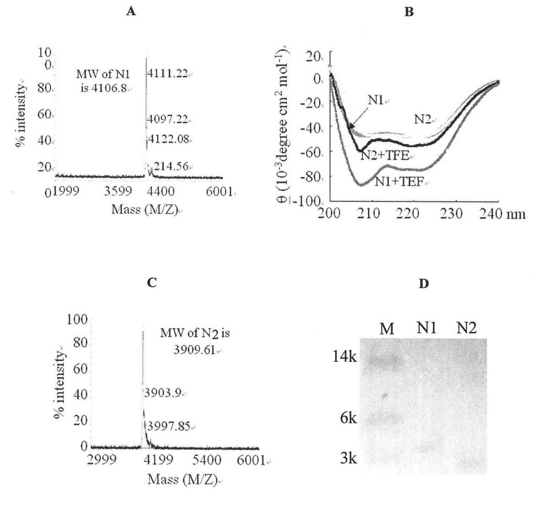
[0095] At the same time, BL21(DE3) bacteria transferred into the empty vector PGEX-6p-1-N2 were used as a control, and no protein was obtained. The SDS-PAGE gel electrophoresis results of N1 and N2 are as follows figure 2 D.
[0096] 2. Analysis of the properties of new peptides
[0097] Circular Dichroism (CD) experiment:
[0098] Method: Adjust the concentration of purified N1 and N2 to 10μM, perform circular dichroism analysis with Jasco J-715 spectrophotometer, operating temperature at 25℃, optical path at 0.1cm, recording wavelength range from 195-235nm;
[0099] The result is figure 2 : Both N1 and N2 have two troughs at 208nm and 222nm, indicating a standard α-helical structure.
[0100] Mass spectrometry (MS):
[0101] Method: The purified N1 and N2 were replaced with a 20mM pH8.0 Tris-HCl buffer with a 3K ultrafiltration tube, and their protein molecular weights were analyzed by Bruker Daltonics Biflex III MALDI-TOF mass spectrometer.
[0102] The result is figure 2 : Obvious main peaks can be seen in the figure, and the results show that the molecular masses of N1 and N2 are 4106.08 Da and 3909.61 Da, respectively.
[0103] 3. Antiviral activity of new peptides
[0104] (1) Anti-infective activity of new peptides against NDV virus
[0105] 1. Cell experiment
[0106] (1) Infection model:
[0107] Increased value of CEF cytotoxicity: Inoculate virus stock in the amount of 1/10 of the cell bottle culture medium. When the cytopathic effect (formation of large syncytia) reaches 75% (usually 48 hours), the virus is harvested by repeated freezing and thawing, that is, the cell bottle is placed Freeze at -20°C for 2 hours and then place it at room temperature until partially thawed. At this time, shake the cell bottle horizontally to detach the adherent cells, and place it again at -20°C for freezing. Repeat this 3 times to release the virus from the cells. Store the cytotoxicity at -20℃ or -70℃.
[0108] Toxicity determination: Dilute the cytotoxicity obtained in step (1) twice to obtain a cytotoxic suspension; culture CEF cells to a monolayer in a 96-well plate, add the cytotoxic suspension to each well, 50μL/well, do 12 One dilution and eight replicates, placed in a carbon dioxide incubator at 37°C for 60 hours, observe the cytopathic lesions and record the fusion rate of the lesions (the fusion rate of lesions is the ratio of the diseased cells forming syncytia to all cells, syncytia is multiple After the fusion of the cell membranes, huge cells are formed, with many nuclei crowded together), and finally the TCID 50 (Virus titer that forms cytopathic disease), repeat 3 times. The results show that the virus TCID of NDV 50 The potency is 10 8.
[0109] Cytopathic effect (CPE) is a cytopathic effect, which refers to the degeneration of cells that occurs after a virus infects tissue culture cells. This effect can be used to quantify the virus. Virus infection forms cytopathies, which are common syncytia (that is, large cells that gather together to form multinuclei) and plaques (cells fall off to form plaques). The disease fusion rate is the ratio of diseased cells to all cells.
[0110] The cell model shows that after the virus stock solution is diluted 1:50,000 times, a 48-well cell plate is inoculated with 50 μL/well and another 50 μL/well culture medium. After 48 hours of culture, the cell plate will show 100 TCIDs. 50 The unit of plaque, this is the virus standard reference amount for the subsequent steps.
[0111] In the protein antiviral inhibition test, IC is often used 50 , Which is the ratio of diseased cells (such as syncytia) to all cells (that is, the fusion rate of diseased) is the concentration corresponding to the protein when 50%, namely 50% inhibitionconstraction, IC 50.
[0112] (2) Inhibition experiment
[0113] Dilute the virus stock solution 1:50,000 times and then inoculate a 48-well cell plate at 50μl/well, add another 50μL/well culture medium, and add protein solutions of different concentrations (50μl/well) at 37℃, CO 2 After culturing in the incubator for 48 hours, stain with 0.5% crystal violet for 3-5 minutes, wash with water and check under microscope. Take five fields of view per well and calculate TCID 50. The virus stock solution used is the same as the virus stock solution used when preparing the infection model (same batch).
[0114] The results show that the antiviral activity of N1 against NDV is IC 50 =0.5μM, the antiviral activity of N2 against NDV is IC 50 =1.0μM, that is, N1 with a concentration of 0.5μM or N2 with a concentration of 1.0μM can effectively inhibit cytopathy, and the ratio of diseased cells to all cells (lesion fusion rate) is close to 50% (the cytopathic situation in the infection model Recorded as 100%); when the N1 and N2 protein concentrations reached 5μM, more than 90% of the cytopathic changes were inhibited.
[0115] 2. Chicken embryo experiment
[0116] (1) Infection model:
[0117] Proliferation of chicken embryo virus: 9-10 day-old chicken embryos are inoculated with 10 μL of virus stock solution (10 6 TCID 50 Unit), collect the allantoic fluid (generally about 35-40 hours) after culturing until the chicken embryo dies, and store it at -20°C or -70°C. Bleeding can be seen in the infected chicken embryo.
[0118] Virulence determination: 9-10 day-old chicken embryos were inoculated with virus stock through the allantoic cavity, and each chicken embryo was inoculated with 10μL virus solution (the amount of virus contained in 10μL virus solution was 10 6 TCID 50 Unit). After the chicken embryo died, the allantoic fluid was collected to determine the blood coagulation titer, the disease of the chicken embryo was observed and the time of death was recorded. As a result, the chicken embryo showed systemic hemorrhagic lesions (the degree of hemorrhagic lesions was +++), the death time of the chicken embryo was 35 hours, and the blood clotting titer was 2 8 Considering that the virus is often infected with high titers in actual production, this virus dilution is used in subsequent experiments. Virus-induced chicken embryo lesions are characterized by hemorrhage (NDV), which can be observed with the naked eye and distinguish between normal and infected conditions.
[0119] (2) Inhibition experiment
[0120] Inoculate the allantoic cavity of 9-10 day-old chicken embryos, and each chicken embryo is inoculated with 10 μL virus solution (the amount of virus in 10 μL virus solution is 10 6 TCID 50 Unit), and add different amounts of protein (0.1mg/bird, 1mg/bird), and collect allantoic fluid to determine the hemagglutination titer and observe the lesions after the chicken embryos die.
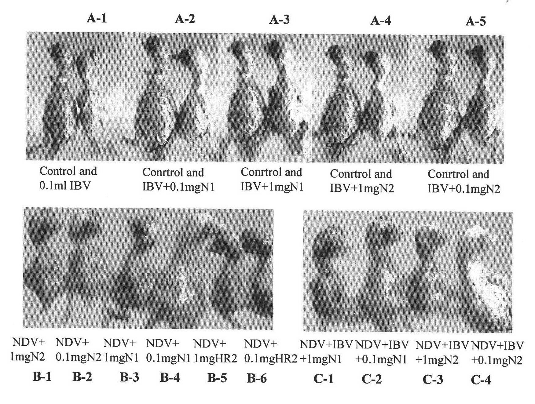
[0121] The results show that 0.1 mg of N1 can effectively inhibit the lesions of NDV infected chicken embryos (ie, the chicken embryos do not bleed, and the degree of bleeding lesions is -), and the chicken embryos can survive up to 72 hours ( image 3 Medium B-4); 1mg of N1 affects the development of chicken embryos, thereby aggravating the toxic effect of NDV (highly virulent) on chicken embryos, which is similar to the disease of chicken embryos in the infection model (the degree of hemorrhagic disease is +++, image 3 B-3). 0.1mg of N2 can effectively inhibit NDV-infected chicken embryo lesions (that is, the chicken embryo does not bleed, and the degree of bleeding lesions is -), but the chicken embryo can only survive up to 45 hours ( image 3 Medium B-2); 1.0 mg of N2 can effectively inhibit NDV-infected chicken embryo lesions (that is, the degree of hemorrhagic lesions is +), but chicken embryos can only survive up to 40 hours ( image 3 B-1).
[0122] The anti-NDV activity results of different doses of protein (polypeptide) are shown in Table 1A.
[0123]
[0124] At the same time, the expression product of BL21(DE3) bacteria transferred into the empty vector PGEX-6p-1-N2 was used for the above experiment photos, and as a result, no antiviral activity was detected.
[0125] At the same time, the expression product of BL21(DE3) bacteria transferred into the empty vector PGEX-6p-1-N1 was used for the above experiment photos, and as a result, no antiviral activity was detected.
[0126] (2) Anti-infective activity of new peptides against IBV virus:
[0127] 1. Cell experiment
[0128] (1) Infection model:
[0129] The proliferation of CEF cytotoxicity: consistent with the experiment (1).
[0130] Toxicity determination: Dilute the cytotoxicity obtained in step (1) twice to obtain a cytotoxic suspension; culture CEF cells to a monolayer in a 96-well plate, add the cytotoxic suspension to each well, 50μL/well, do 12 One dilution and eight repetitions. After placing them in a carbon dioxide incubator at 37°C for 48 hours, observe the cytopathic changes and record the fusion rate of the lesions (the fusion rate of lesions is the ratio of the diseased cells that form syncytia to all cells, and syncytia is multiple After the cell membrane is fused, a huge cell is formed, with many nuclei crowded together), and finally the TCID is calculated 50 (The titer of virus that forms cytopathic disease), repeat 3 times. The results show that the virus TCID of IBV 50 The potency is 10 -7.
[0131] The cell model shows that after the virus stock solution is diluted 1:5000 times, a 48-well cell plate is inoculated at 50 μL/well, and another 50 μL/well culture medium is added. After 48 hours of culture, the cell plate will show 100 TCIDs. 50 The unit of plaque, this is the virus standard reference amount for the subsequent steps.
[0132] (2) Inhibition experiment
[0133] Dilute the virus stock solution 1:5000 times and then inoculate a 48-well cell plate. Add 50μL/well and add another 50μL/well culture solution. At the same time, add different concentrations of protein solution (50μl/well), 37℃, CO 2 After culturing in the incubator for 48 hours, stain with 0.5% crystal violet for 3-5 minutes, wash with water and check with microscope. Take five fields of view per well and calculate TCID 50. The virus stock solution used is the same as the virus stock solution used when preparing the infection model (same batch).
[0134] The results show that the antiviral activity of N1 against IBV is IC 50 =0.8μM, the antiviral activity of N2 against IBV is IC 50 =1.0μM, that is, N1 with a concentration of 0.8μM or N2 with a concentration of 1.0μM can effectively inhibit cytopathy, and the ratio of diseased cells to all cells (fusion rate) is close to 50% (the cytopathic situation in the infection model is recorded 100%); when the N1 and N2 protein concentrations reached 5μM, more than 90% of the cytopathic changes were suppressed.
[0135] 2. Chicken embryo experiment
[0136] (1) Infection model:
[0137] Proliferation of chicken embryo virus: 9-10 day-old chicken embryos are inoculated with 100μL of virus stock solution per embryo through the allantoic cavity, and allantoic fluid is collected 72 hours after the inoculation of chicken embryos, and stored at -20℃ or -70℃. IBV infected chickens The embryo does not die, but the chicken embryo is obviously smaller (dwarf embryo).
[0138] Virulence determination: 9-10 day old chicken embryos are inoculated with virus solution through the allantoic cavity, and each chicken embryo is inoculated with 100μL (100μL virus solution contains 10 6 TCID 50 Unit) Virus solution, collect allantoic fluid 72 hours after inoculation of chicken embryos, observe the disease of chicken embryos. As a result, the chicken embryo showed a smaller disease state (the length of the main body is about 2/3 of the volume of the normal control chicken embryo, that is, the degree of dwarf disease is +++), considering the high titer of the virus in actual production In the case of infection, this virus dilution was used in all subsequent experiments.
[0139] Virus-induced chicken embryo lesions are mostly characterized by small body (IBV), which can be observed with the naked eye and distinguish between normal and infected conditions.
[0140] (2) Inhibition experiment
[0141] Inoculate the allantoic cavity of 9-10 day-old chicken embryos, each chicken embryo is inoculated with 100μL virus solution (100μL virus solution contains 10 6 TCID 50 Unit), and add different amounts of protein (0.1mg/head, 1mg/head), collect allantoic fluid and observe the lesion after 72 hours of culture.
[0142] The results of the anti-IBV activity of each polypeptide are shown in Table 1B.
[0143] The results show that 0.1 mg of N1 can effectively inhibit the pathological changes of IBV infected chicken embryos (the length of the diseased chicken embryo is 4/5 of the normal chicken embryo, that is, the degree of dwarf disease is ++, see image 3 Medium A-2); 1mg of N1 can almost completely stop the virus from attacking chicken embryos (that is, no dwarf embryos appear at all, and the degree of dwarf disease is -, see image 3 Middle A-3). 0.1mg of N2 can also effectively inhibit the lesions of IBV infected chicken embryos (the length of diseased chicken embryos is 5/6 of that of normal chicken embryos, that is, the degree of dwarf disease is +, see image 3 Middle A-5); 1mg of N2 inhibits lesions slightly better than 0.1mg of N2 (the length of the diseased chicken embryo is 5/6 of the normal chicken embryo, that is, the degree of dwarf disease is +, see image 3 Medium A-4), but considering the impact of a large amount of heterologous protein on the body and future price issues, 0.1mg is more advantageous. among them image 3 "0.1ml IBV" in A-1 refers to infected chicken embryos with the following control bacteria expression products added. image 3 The middle control means normal chicken embryos that have not been infected with the virus.
[0144] At the same time, the expression product of BL21(DE3) bacteria transferred into the empty vector PGEX-6p-1-N2 was used for the above-mentioned experimental photos. As a result, no antiviral activity was detected.
[0145] At the same time, the expression product of BL21(DE3) bacteria transferred into the empty vector PGEX-6p-1-N1 was used for the above experiment photos, and as a result, no antiviral activity was detected.
[0146] (3) Anti-infective activity of the new peptides against MDV virus:
[0147] 1. Cell experiment
[0148] (1) Infection model:
[0149] The proliferation of CEF cytotoxicity: consistent with the method described in experiment (1).
[0150] Toxicity test: Dilute the cytotoxicity obtained in step (1) twice to obtain a cytotoxic suspension; culture CEF cells to a monolayer in a 96-well plate, add the cytotoxic suspension to each well, 50μL/well, do 12 One dilution and eight replicates, placed in a carbon dioxide incubator at 37°C for 72 hours, observe the cytopathic changes and record the fusion rate of the lesions, and finally calculate the TCID 50 (The titer of virus that forms cytopathic disease), repeat 3 times. The results show that the virus TCID of MDV 50 The potency is 10 -5.
[0151] The cell model shows that after the virus stock solution is diluted 1:50 times, a 48-well cell plate is inoculated with 50 μL/well and another 50 μL/well culture medium. After 48 hours of culture, the cell plate will have 100 TCIDs. 50 The unit of plaque, this is the virus standard reference amount for the subsequent steps.
[0152] (2) Inhibition experiment
[0153] Dilute the virus stock solution 1:50 times and then inoculate a 48-well cell plate at 50μL/well, add another 50μL/well culture medium, and add different concentrations of protein solution (50μl/well), 37℃, CO 2 After culturing in the incubator for 48 hours, stain with 0.5% crystal violet for 3-5 minutes, wash with water and check with microscope. Take five fields of view per well and calculate TCID 50. The virus stock solution used is the same as the virus stock solution used when preparing the infection model (same batch).
[0154] The results show that the antiviral activity of N1 against MDV is IC 50 =1.5μM, that is, N1 at a concentration of 1.5μM can effectively inhibit cytopathic changes, and the ratio of diseased cells to all cells (lesion fusion rate) is close to 50% (the cytopathic situation in the infection model is recorded as 100%); When the N1 and N2 protein concentration reached 5μM, more than 90% of the cytopathic changes were suppressed.
[0155] 2. Chicken embryo experiment
[0156] (1) Infection model:
[0157] The proliferation of chicken embryo poison: consistent with the method described in experiment (1).
[0158] Virulence determination: 9-10 day old chicken embryos are inoculated with different concentrations of virus stock through the yolk sac, and each chicken embryo is inoculated with 10μL (the amount of virus contained in 10μL virus solution is 10 3 TCID 50 Unit) Virus solution, 8-10 days later, when the chicken embryos die, take out the chorioallantoic membrane of the chicken embryos, observe the tumor formation under a microscope and take pictures. The experiment is repeated 3 times. As a result, clear tumors on the chick embryo chorioallantoic membrane were seen in the chicken embryo (50 gray pox tumors/field of view on average at 40x focal length). After that, the virus titer was used in the experiment.
[0159] The disease of chicken embryo caused by MDV virus is characterized by inflammatory reaction such as immune cell infiltration or tumor formation, which can be observed under a microscope to distinguish between normal and infected conditions.
[0160] (2) Inhibition experiment
[0161] Inoculate the allantoic cavity of 9-10 day-old chicken embryos, and each chicken embryo is inoculated with 10 μL virus solution (the amount of virus in 10 μL virus solution is 10 3 TCID 50 Unit), and add different amounts of protein (0.1mg/bird, 1mg/bird), 8-10 days later when the chicken embryo dies, take out the chick embryo chorioallantoic membrane, observe the tumor formation, the experiment is repeated 3 times.
[0162] The results show that 0.1 mg of N1 can effectively inhibit the formation of chick embryo chorioallantoic membrane tumors (that is, there is no tumor in each field of view at 40 times the focal length). Figure 4 It is the result of MDV-chicken chorioallantoic membrane tumor lesion. The left picture shows the normal chicken chorioallantoic membrane under the microscope, the middle picture shows the tumor formation of the chick embryo chorioallantoic membrane, and the right picture shows the tumor formation inhibited by 0.1mg of N1.
[0163] At the same time, the expression product of BL21(DE3) bacteria transferred into the empty vector PGEX-6p-1-N2 was used for the above-mentioned experimental photos. As a result, no antiviral activity was detected.
[0164] At the same time, the expression product of BL21(DE3) bacteria transferred into the empty vector PGEX-6p-1-N1 was used for the above experiment photos, and no antiviral activity was detected.
[0165] (4) Anti-infective activity of the new peptides against IBV virus and NDV virus:
[0166] 1. Chicken embryo experiment
[0167] (1) Infection model:
[0168] Propagation of chicken embryo virus: respectively proliferate IBV virus and NDV virus, the method is the same as that described in experiment (1).
[0169] Inoculate the mixed virus of IBV and NDV in the following amounts: (1) The inoculation amount of NDV is 10μL (the amount of virus contained in 10μL virus solution is 10 6 TCID 50 ), the inoculation volume of IBV is 100 μL (the amount of virus contained in 100 μL virus solution is 10 6 TCID 50 ); denoted as 10 6 TCID 50 (Original poison 10μL): 10 6 TCID 50 (Original poison 100μL) group;
[0170] (2) The amount of NDV inoculated is 10 μL (the amount of virus contained in 10 μL virus solution is 10 6 TCID 50 ), the inoculation volume of IBV is 100 μL (the amount of virus contained in 0.1 μL virus solution is 10 3 TCID 50 , Namely the original poison 1μL diluted 10 3 After doubling, take 100μL to challenge); denote as 10 6 TCID 50 (Original poison 10μL): 10 3 TCID 50 (Original poison 100μL) group;
[0171] (3) The amount of NDV inoculated is 10 μL (the amount of virus contained in 10 μL virus solution is 10 6 TCID 50 ), the inoculation volume of IBV is 100μL (10 -4 The amount of virus contained in μL virus solution is 10 0 TCID 50 , Namely the original poison 1μL diluted 10 6 After doubling, take 100μL to challenge); denote as 10 6 TCID 50 (Original poison 10μL): 10 0 TCID 50 (Original poison 100μL) group.
[0172] Toxicity determination: 9-10 day old chicken embryos were inoculated with mixed virus solution through the allantoic cavity according to the above amount. After the chicken embryos died, the allantoic fluid was collected to determine the hemagglutination titer, observe the chicken embryo lesions and record the time of death. As a result, 10 6 TCID 50 (Original poison 10μL): 10 6 TCID 50 (Original virus 100μL) group, chicken embryos have obvious systemic hemorrhage lesions (the degree of bleeding lesions is +++) and dwarf lesions (the length of diseased chicken embryos is about 9/10 of the length of normal chicken embryos, and the degree of dwarf lesions is +++) , The chicken embryo dead time is 40 hours (the bleeding symptoms can be clearly observed with the naked eye, while the difference in dwarf lesions is not obvious), and the blood clotting titer is 2 7 Considering the high-titer virus infection in actual production, this virus dilution and ratio are used in all subsequent experiments.
[0173] (2) Inhibition experiment
[0174] According to the preparation infection model in 10 6 TCID 50 (Original poison 10μL): 10 6 TCID 50 (100μL of the original virus) group, inoculate the mixed virus of NDV and IBV into the allantoic cavity of 9-10 day old chicken embryos, each chicken embryo is inoculated with 110μL virus solution, and different amounts of protein (0.1mg/bird, 1mg/head), after the chicken embryo died, the allantoic fluid was collected to determine the hemagglutination titer, the disease of the chicken embryo was observed and the death time was recorded.
[0175] The results of the anti-NDV and IBV mixed infection activity of each polypeptide are shown in Table 1C.
[0176] Table 1C
[0177]
[0178] The results showed that N1 and N2 polypeptides not only have strong inhibitory ability on single virus infection, but also have strong inhibitory ability on mixed virus infection. 0.1mg of N1 can effectively inhibit the disease of chicken embryos (chicken embryos) mixed with NDV and IBV viruses. The degree of hemorrhagic lesions is +, and the degree of dwarfism is not obvious, which is similar to that of normal chicken embryos (that is, the degree of dwarfing lesions is -), but the chicken embryos can only survive up to 45 hours ( image 3 Medium C-2); 1mg of N1 also showed the side effects of polypeptide, which is similar to the degree of disease of infected chicken embryo, the degree of bleeding and dwarf disease are both +++ ( image 3 C-1). 0.1mg of N2 can also effectively inhibit the disease of chicken embryos mixed with NDV and IBV virus (that is, there is no hemorrhage of chicken embryos and no dwarf embryos, the degree of hemorrhagic disease is -, the degree of dwarf disease is -), and the chicken embryo can survive to 72 hour( image 3 C-4). 1mg of N2 can also effectively inhibit the lesions of mixed-infected chicken embryos (ie, chicken embryo bleeding and dwarf lesions are reduced, the degree of bleeding lesions and dwarf lesions are both +, and the length of the diseased chicken embryo is about 9.5/ the length of the normal chicken embryo. 10, that is, the degree of dwarf disease is +), chicken embryos can survive up to 40 hours, but the effect is not as good as 0.1 mg of N1 ( image 3 C-3).
[0179] At the same time, the expression product of BL21(DE3) bacteria transferred into the empty vector PGEX-6p-1-N2 was used for the above-mentioned experimental photos. As a result, no antiviral activity was detected.
[0180] At the same time, the expression product of BL21(DE3) bacteria transferred into the empty vector PGEX-6p-1-N1 was used for the above experiment photos, and as a result, no antiviral activity was detected.
[0181] The above results show that 0.1mg N1 has obvious activity against NDV and 0.1mg N2 against IBV and mixed infections. However, if the concentration is too large (such as 1mg), it will affect the development of chicken embryos, thereby aggravating the effects of NDV (highly virulent) on chicken embryos. The poisonous effect of saccharine (see Figure C-1 and B-3). The length of the newly constructed polypeptide is similar to the respective HR2 regions of the two viruses (both are about 30 amino acids), but the antiviral activity is more effective (cell-level IC 50 Less than 1.0 μM unit, the level of chicken embryo is about 0.1 mg unit), and it is also effective in anti-mixed infection.

Example Embodiment

[0182] Example 2. Preparation and functional test of control polypeptide
[0183] 1. Antiviral activity of IBV HR2 polypeptide, NDV HR2 polypeptide and MDV HR2 polypeptide
[0184] (1) Preparation of each polypeptide
[0185] 1. Gene cloning and protein expression and purification: the following PCR products were digested and recovered and cloned into the PGEX-6p-1 vector (purchased from Amasia, product catalog number P0048), and the positive clones were transformed into BL21 (DE3) ) Bacteria (purchased from Beijing Kangwei Century Company, product catalog number CW0809) for protein expression, ultrasonically lysed the expressed cells and centrifuged to collect the supernatant. The supernatant was passed through a Glutathione-Sepharose 4B affinity chromatography column equilibrated with PBS ( (Purchased from Oriental Technology Company), then wash the affinity column with PBS for at least 10 column volumes, and then use at least 3 column volumes of reduced glutathione solution (10mmol/L reduced glutathione, 50mmol/L Tris·HCl, pH 8.0) Elution, collect the eluate to obtain the GST fusion protein, then concentrate it with an ultrafiltration tube with a cut-off molecular weight of 5K protein, and then change to enzyme digestion buffer (50mmol/L Tris·HCl (pH 7.0), 150mmol/L) NaCl, 1mmol/L DTT, 1mmol/L EDTA, pH 8.0), add excess GST-3C protease (purchased from Dongfang Technology Co., Ltd.), digest for 16 hours at 4°C, concentrate and then change to PBS buffer, use Glutathione-Sepharose 4B The affinity chromatography column removes the digested GST and GST-3C by adding an appropriate amount of Glutathione-Sepharose 4B affinity filler equilibrated with PBS, shaking gently to mix, and constantly shaking in the middle to prevent filler precipitation. The effect is about After 1h (or a little longer time), centrifuge at 500rpm for 15min, and gently aspirate the supernatant to obtain the purified target protein. Use a 3K ultrafiltration tube to concentrate to a concentration of about 3mg/mL. Perform 15% SDS- on the purified target protein. PAGE detection showed that the target protein with higher purity was obtained. Store the target protein frozen at -80°C.
[0186] When amplifying the HR2 polypeptide encoding gene of IBV, refer to the IBV gS gene sequence (GeneBank No.: AAK27168), and use the bridge PCR method to amplify the HR2 gene. The amino acid sequence of the HR2 polypeptide is: DFDKFNYTVP I LD IG SE I DR IQ GV I QG LN DS L ID LE TL S IL KT Y; (underlined are the a, d, e positions of amino acids); the base sequence of its coding gene is as follows: gac ttc gac aaa ttc aac tac aca gta cct ata cttgac att gat agt gaa att gat cgt att caa ggc gtt ata cag ggt ctt aat gac tctcta ata gac ctt gaa aaa ctt tcc atc ctg aaa act tat; primer design method: 1 and 3 primer sequences are the partial fragments in the same direction as the target gene, 1 primer contains BamHI restriction site (underlined) ; And primers 2, 4 in turn are partial fragments complementary to the target gene and have a 15-18 bp bridging region with primers A and 3 in turn, primer 4 contains the XhoI restriction site (underlined), and the restriction site is preceded by Stop codon. The length of each primer must be less than 60bp, and the G+C content of all primers and the bridging area must be controlled at about 50%. The primer sequence is as follows (5'-3'): 1, gcc gga tcc gac ttc gac aaa ttc aac tac act gta cca atc ​​ctg gac; 2, cgccct gaa tac gat caa tct cgc tgt caa tgt cca gga ttg gta c; 3. at cgt att cagggc gtt atc cag ggt ctg aac gac tct ctg atc gac ctt g; 4, gcg ctc gag tca ataagt ttt cag gat gga cag ctt ctc aag gtc gat cag ag
[0187] When amplifying the gene encoding the HR2 polypeptide of NDV, refer to the sequence of the gF gene of NDV (GenBank Accession No.AF079172), and amplify the HR2 gene by bridge PCR. The amino acid sequence of the HR2 polypeptide is: The amino acid sequence of the HR2 polypeptide of NDV is: L GN VN NS I SN AL DK L EE SN SK L DK VN VK L TG TS ALI (underlined are the a, d, e positions of amino acids); the base sequence is as follows: ctt ggt aat gtc aac aac tcg ata agt aat gctttg gat aag tta gag gaa agc aat agc aaa ctt gac aaa gtc aat gtc aag ctg accggc act tct gct ctg att; the primer sequence is as follows (5′-3′): 1, agc gga tcc ctt ggt aat gtcaac aac agc atc agc aac gct ctg gat; 2, gtc aag ctt gct gtt gct ttc ctc taactt atc cag agc gtt gct ga; 3, ac agc aag ctt gac aaa gtc aac gtc aag ctt actggc ac; 4, cag ctc gag tca aat cag agc aga agt gcc agt aag ctt gac;
[0188] When amplifying the HR1 polypeptide coding gene of MDV, first refer to the MDV gH gene sequence (GeneBank No.: GeneBank No.: AY129989), and design primers (5′-3′): upstream primer: ata gga tcc atg ggt cttccc ggt agt at, downstream primer: cgc gaa ttc tta aag atc gtc gta cag gc. Then use the gH plasmid as a template to amplify the HR1 gene by ordinary PCR. The amino acid sequence of the HR1 polypeptide is: M NA SD ME I KS YI NM I ES VE ES S N YD F (underlined are the a, d, e positions of amino acids); the base sequence is as follows: atgaac gca tcg gat atg gaa att aaa tca tat att aat gtg atc gaa agt gta gag gaatca tcc aat tat gat ttc; the primer sequence is as follows (5 '-3'): Upstream primer: caa gga tcc cat atgaac gca tcg gat; downstream primer: gcg ctc gag tca gaa atc ​​ata att gga tga.
[0189] (2) Antiviral infection activity of IBV HR2 polypeptide:
[0190] 1. Cell experiment
[0191] The method is the same as that described in Experiment 3 (2) 1 (2) in Example 1. The result shows that the IC of IBV HR2 polypeptide 50 The titer is 16μM; the protein has no inhibitory activity against other viruses.
[0192] 2. Chicken embryo experiment
[0193] The method is the same as that described in Experiment 3 (2) 2 (2) in Example 1. The results show that 1 mg of IBV HR2 polypeptide can effectively inhibit the pathological changes of IBV-infected chicken embryos (pathological degree is +), and the effect is similar to 0.1 mg of N2, but the inhibitory activity of 0.1 mg unit of polypeptide is not obvious.
[0194] (3) Antiviral infection activity of HR2 polypeptide of NDV:
[0195] 1. Cell experiment
[0196] The method is the same as that described in Experiment 3 (1) 1 (2) in Example 1. The results showed that the IC of NDV's HR2 polypeptide 50 The titer is 15μM; the protein has no inhibitory activity against other viruses;
[0197] 2. Chicken embryo experiment
[0198] The method is the same as that described in Experiment 3 (1) 2 (2) in Example 1. The results show that 0.1 mg of NDV's HR2 polypeptide can effectively inhibit the lesions of NDV infected chicken embryos (the degree of lesions is ++) ( image 3 Medium B-6), but chicken embryos can only survive up to 40 hours, and the side effects of peptides in 1 mg units are more obvious ( image 3 B-5).
[0199] (4) Antiviral infection activity of HR1 polypeptide of MDV:
[0200] 1. Cell experiment
[0201] The method is the same as that described in Experiment 3 (3) 1 (2) in Example 1. The results show that IC 50 The potency is 10μM.
[0202] 2. Chicken embryo experiment
[0203] The method is the same as that described in Experiment 3 (3) 2 (2) in Example 1. The results showed that 1 mg of MDV HR1 polypeptide effectively inhibited the formation of tumors on the chorioallantoic membrane (that is, there was no tumor on the chick embryo chorioallantoic membrane), but the effect was similar to that of 0.1 mg of N1, and 0.1 mg of the polypeptide could not completely inhibit Tumor formation (inhibition rate of about 50%).
[0204] 2. Antiviral infection activity of the tandem polypeptide (I-L-N) formed by the HR2 polypeptide of IBV and the HR2 polypeptide of NDV
[0205] (1) Preparation of tandem polypeptide (I-L-N)
[0206] The amino acid sequence of the tandem polypeptide (I-L-N) is: DFDKFNYTVP I LD IG SE I DR IQ GV I QG LN DS L ID LE TL S IL KT YSGGRGG L GN VN NS I SN AL DK L EE SN SK L DK VN VK L TG TS ALI (underlined are the a, d, e positions of amino acids); the base sequence is as follows: gac ttc gac aaa ttcaac tac aca gta cct ata ctt gac att gat agt gaa att gat cgt att caa ggc gttata cag ggt ctt aat gac tct cta ata gac ctt gaa aaa ctt tcc atc ctg aaa acttat tct ggt gga cga gga ggt ctt ggt aat gtc aac aac tcg ata agt aat gct ttggat aag tta gag gaa agc aat agc aaa ctt gac aaa ct gtc act gt g att;
[0207] Use IBV-HR2 as template, PCR primers bv1 and bvnew1, synthesize IBVHR2-linker (first round); at the same time, use NDV-HR2 (same as in experiment two, (1), 1) as template, PCR primers bv2 and bvnew2, synthesize linker-NDVHR2 (first round); then use IBVHR2-linker and -linkerNDVHR2 as templates, primers bv1 and bvnew3, and finally synthesize IBVHR2-linker-NDVHR2. The primer sequence is as follows (5'-3'): bv1: gccgga tcc gac ttc gac aaa ttc aac tac act gta cca atc ​​ctg gac; bv2: tct ggt ggacga gga ggt ctt ggt aat gtc aac aac; bvnew1: acc tcc tcg tcc acc aga ata agtttt cag gat gga; bvnew2: cag ctc gag tca aat cag agc aga agt gcc; bvnew3: cag ctc gag tca aat cag agc aga agt gcc a.
[0208] The method of expression and purification of the I-L-N polypeptide is the same as the method of expression and purification of the protein described in Example 1.
[0209] (2) Structural research of tandem peptides:
[0210] The ILN peptide was analyzed by circular dichroism and molecular sieve analysis (automatically performed by the instrument). The results showed that the ILN peptide was a monomer, and the secondary structure was similar to the structure of each constituent unit (NDVHR2 and IBVHR2), indicating that NDVHR2 and IBVHR2 did not bind to each other .
[0211] (3) Anti-infective activity of tandem peptides against NDV virus:
[0212] 1. Cell experiment
[0213] The method is the same as that described in Experiment 3 (1) 1 (2) in Example 1. The results show that the IC of I-L-N polypeptide 50 The potency is 10μM.
[0214] 2. Chicken embryo experiment
[0215] The method is the same as that described in Experiment 3 (1) 2 (2) in Example 1. The results show that 0.1 mg of I-L-N can also effectively inhibit the lesions of chicken embryos infected by NDV (the degree of disease is +), but the chicken embryos can only survive up to 40 hours; 1 mg of I-L-N has obvious side effects.
[0216] (4) Anti-infective activity of tandem peptides against IBV virus:
[0217] 1. Cell experiment
[0218] The method is the same as that described in Experiment 3 (2) 1 (2) in Example 1. The results show that the IC of I-L-N polypeptide 50 The potency is 15 μM.
[0219] 2. Chicken embryo experiment
[0220] The method is the same as that described in Experiment 3 (2) 2 (2) in Example 1. The results show that 1 mg of I-L-N can also effectively inhibit the pathological changes of IBV-infected chicken embryos (the degree of disease is ++), and the effect is similar to that of 0.1 mg of N1; 1.0 mg of I-L-N has no obvious inhibitory effect.
[0221] (5) Anti-infective activity of tandem peptides against IBV virus and NDV virus:
[0222] The method is the same as that described in Experiment 3 (4) 1 (2) in Example 1. The results showed that 0.1 mg of ILN can effectively inhibit the lesions of virus mixed infection of chicken embryos (both hemorrhage and dwarfing lesions are +), and the effect is similar to 1.0 mg of N2, chicken embryos can survive up to 40 hours, and 1.0 mg of ILN also It can effectively inhibit the lesions of mixed virus infection of chicken embryos (both hemorrhage and dwarfing lesions are +), but the chicken embryos can only survive up to 35 hours.
[0223] 3. Anti-infective activity of modified tandem peptides
[0224] (1) Modification of tandem peptides
[0225] Two lysines (K) and two glutamic acids (E) are connected in series (ie EEKK) to modify the amino terminus of I-L-N, that is, EK-I-L-N by genetic engineering. The amino acid sequence is: EEKKDFDKFNYTVP I LD IG SE I DR IQ GV I QG LN DS L ID LE TL S IL KT YSGGRGG L GN VN NS I SN AL DK L EE SN SK L DK VN VK L TG TS ALI (underlined are the a, d, e positions of amino acids); the base sequence is as follows: gag gag aag aaggac ttc gac aaa ttc aac tac aca gta cct ata ctt gac att gat agt gaa att gatcgt att caa ggc gtt ata cag ggt ctt aat gac tct cta ata gac ctt gaa aaa ctttcc atc ctg aaa act tat tct ggt gga cga gga ggt ctt ggt aat gtc aac aac tcgata agt aat gct ttg gat aag tta gag gaa agc ag tta gag gaa agc ag agc agc agc act tct gct ctg att; method: use ILN plasmid (see two, (two), 1) as a template to construct the target gene, the upstream primer is: cgc gga tcc gag gag aag aag aag gac ttc gac aaattc aac, the downstream primer is: cag ctc gag tca aat cag agc aga agt gcc a.
[0226] The expression and purification method of EK-I-L-N is the same as the expression and purification method of the protein described in Example 1.
[0227] (2) Anti-infective activity of the modified tandem polypeptide against NDV virus:
[0228] 1. Cell experiment
[0229] The method is the same as that described in Experiment 3 (1) 1 (2) in Example 1. The results show that the antiviral activity of EK-I-L-N against NDV is IC 50 =5μM.
[0230] 2. Chicken embryo experiment
[0231] The method is the same as that described in Experiment 3 (1) 2 (2) in Example 1. The results show that 0.1 mg of EK-ILN can also effectively inhibit the lesions of chicken embryos infected by NDV (the degree of lesion is +), and the chicken embryos can survive up to 45 hours; 1 mg of EK-ILN can also effectively inhibit the lesions of chicken embryos infected by NDV (The degree of disease is ++), chicken embryos can only survive up to 45 hours.
[0232] (3) Anti-infective activity of modified tandem polypeptide against IBV virus:
[0233] 1. Cell experiment
[0234] The method is the same as that described in Experiment 3 (2) 1 (2) in Example 1. The results show that the antiviral activity of EK-I-L-N against IBV is IC 50 = 6 μM.
[0235] 2. Chicken embryo experiment
[0236] The method is the same as that described in Experiment 3 (2) 2 (2) in Example 1. The results show that 1 mg of EK-ILN can also effectively inhibit the pathological changes of IBV infected chicken embryos (the degree of pathology is +), but the effect is similar to 0.1 mg of N2; 0.1 mg of EK-ILN can also effectively inhibit the pathological changes of IBV infected chicken embryos (The degree of lesion is ++), but the effect is similar to 0.1mg of N1.
[0237] (4) Anti-infective activity of modified tandem polypeptide against IBV virus and NDV virus:
[0238] The method is the same as that described in Experiment 3 (4) 1 (2) in Example 1. The results showed that 0.1 mg and 1 mg of EK-ILN can also effectively inhibit the mixed virus infection of chicken embryo lesions (both hemorrhage and dwarfing lesions are +), and the effect is similar to 1 mg of N2, but chicken embryos can survive to 45. Hours and 40 hours.
[0239] Studies have proved that the HR2 regions of IBV virus and NDV virus do not bind to each other and have no cross antiviral activity, which proves that although the HR regions of different viruses have their own inherent characteristics, their specificity is obvious; the polypeptide expressed in tandem is highly effective against viruses The infection/mixed infection activity proves that there is no mutual interference by the spatial structure; the newly constructed polypeptide modified by E/K has more significant antiviral activity, which proves that the formation of the salt bridge can promote the function of the heptapeptide repeat region.

PUM

PropertyMeasurementUnit
Molecular mass3909.61 ~ 4106.08

Description & Claims & Application Information

We can also present the details of the Description, Claims and Application information to help users get a comprehensive understanding of the technical details of the patent, such as background art, summary of invention, brief description of drawings, description of embodiments, and other original content. On the other hand, users can also determine the specific scope of protection of the technology through the list of claims; as well as understand the changes in the life cycle of the technology with the presentation of the patent timeline. Login to view more.

Similar technology patents

Detection method of real-time voltage vectors in energy feedback unit

InactiveCN101900751AStrong inhibitory ability
Owner:雷富特节能科技(上海)有限公司

Electric automobile charging pile with positioning and voice prompt functions

InactiveCN104967158AStrong inhibitory abilityReduce misuse
Owner:SHAANXI XINWANG NEW ENERGY TECH

High-power harmonic suppression filter based on rectangular waveguide structure

ActiveCN112563705AIn-band insertion loss is smallStrong inhibitory ability
Owner:SOUTHEAST UNIV

High-precision low-voltage voltage/current switching circuit

ActiveCN101694963Alow working voltageStrong inhibitory ability
Owner:MAXIC TECHNOLOGY CORPORATION

Copper-sulfur separation inhibitor, lime-free copper-sulfur flotation separation method and application thereof

PendingCN112604816AStrong inhibitory abilitygood choice
Owner:BEIJING MINING & METALLURGICAL TECH GRP CO LTD +1

Classification and recommendation of technical efficacy words

  • good stability
  • Strong inhibitory ability

Detection method of real-time voltage vectors in energy feedback unit

InactiveCN101900751AStrong inhibitory ability
Owner:雷富特节能科技(上海)有限公司

High-precision low-voltage voltage/current switching circuit

ActiveCN101694963Alow working voltageStrong inhibitory ability
Owner:MAXIC TECHNOLOGY CORPORATION

Electric automobile charging pile with positioning and voice prompt functions

InactiveCN104967158AStrong inhibitory abilityReduce misuse
Owner:SHAANXI XINWANG NEW ENERGY TECH

High-power harmonic suppression filter based on rectangular waveguide structure

ActiveCN112563705AIn-band insertion loss is smallStrong inhibitory ability
Owner:SOUTHEAST UNIV

Copper-sulfur separation inhibitor, lime-free copper-sulfur flotation separation method and application thereof

PendingCN112604816AStrong inhibitory abilitygood choice
Owner:BEIJING MINING & METALLURGICAL TECH GRP CO LTD +1
Why Eureka
Try Eureka