Three-dimensional nano stent of gene delivery system, preparation method and application thereof

A gene delivery system, three-dimensional nanotechnology, applied in the fields of gene transfection, three-dimensional nano-scaffold, and seed cell culture, can solve the problems of difficult to achieve growth factors and so on.

Active Publication Date: 2010-09-15
JIANGSU UNIV
View PDF4 Cites 4 Cited by
  • Summary
  • Abstract
  • Description
  • Claims
  • Application Information

AI Technical Summary

Problems solved by technology

Traditional gene delivery technology, whether it is viral or non-viral, generally only transien

Method used


Image

  • Three-dimensional nano stent of gene delivery system, preparation method and application thereof
  • Three-dimensional nano stent of gene delivery system, preparation method and application thereof
  • Three-dimensional nano stent of gene delivery system, preparation method and application thereof

Examples


Embodiment 1

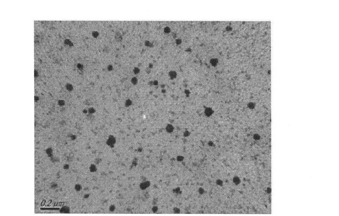
[0017] Example 1: Preparation of calcium phosphate nanoparticles loaded with therapeutic genes

[0018] (1) Preparation: Dissolve 0.1M Igepal CO-520 in 25ml cyclohexane, add 1.36M calcium chloride, stir and mix to form microemulsion 1; dissolve 0.1M Igepal CO-520, 50μl Tis-HCL (pH 7.4) In 25ml of cyclohexane, add disodium hydrogen phosphate and TGF-β1 plasmid, the mass of disodium hydrogen phosphate added is 3.4785 μg, the mass ratio of disodium hydrogen phosphate to plasmid is 0.1:1~10:1, stir to form microemulsion 2. Slowly add microemulsion 2 to microemulsion 1 dropwise, and stir for 10 minutes to form an emulsion of gene-loaded calcium phosphate composite nanoparticles.

[0019] (2) Separation of calcium phosphate composite nanoparticles loaded with genes:

[0020] (a) Pretreatment of chromatography column:

[0021] Take 90g of silicon spheres and add them to 150mL ethanol solution containing 0.336mL of aminopropyltriethoxysilane (APS), 1.5mL of glacial acetic acid, and ...

Embodiment 2

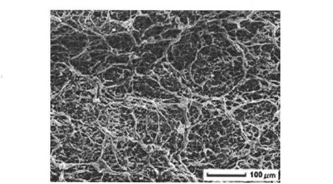
[0026] Embodiment 2: the preparation of a kind of three-dimensional support as contrast

[0027] Get 0.1ml of 1% collagen acetic acid solution, add 0.1ml of 1% chitosan solution, freeze-dry, add 0.25% glutaraldehyde 50 μ L to cross-link, room temperature overnight, freeze-dry, PBS wash three times, freeze-dry, add fibronectin ( FN) 1 μg, to obtain a three-dimensional scaffold that does not contain calcium phosphate nanoparticles loaded with therapeutic genes, the electron micrograph of which is shown in figure 2 .

Embodiment 3

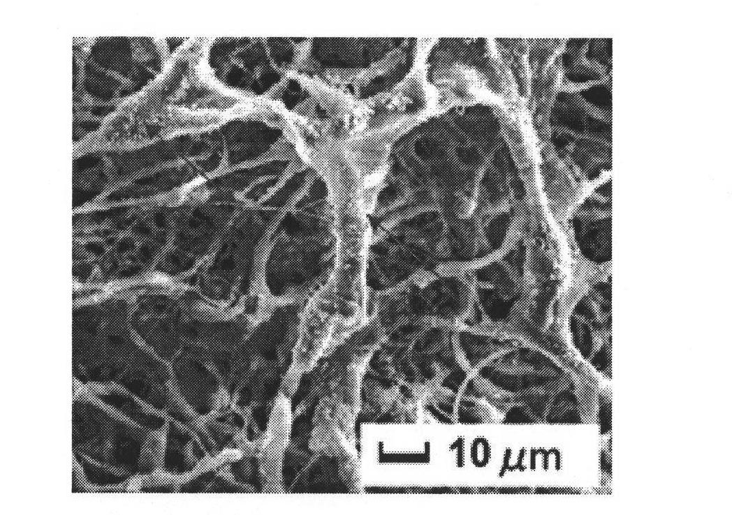
[0028] Embodiment 3: Preparation of the three-dimensional nano-scaffold of the present invention

[0029]Take 0.1ml of 1% collagen acetic acid solution, add 0.1ml of 1% chitosan solution, freeze-dry, add 0.25% glutaraldehyde 50 μ L for cross-linking, room temperature overnight, freeze-dry, wash three times with PBS, 20min / each time, cell culture medium ( Dulbecco'sModified Eagle Media, DMEM) soaked overnight; PBS washed three times, 20min / every time, added the gene-loaded calcium phosphate composite nanoparticles 1 μg and FN1 μg prepared in Example 1, freeze-dried to obtain the three-dimensional nano-scaffold of the present invention, Its electron microscope photo see image 3 .


PUM

PropertyMeasurementUnit
Particle sizeaaaaaaaaaa

Abstract

The invention discloses a three-dimensional nano stent of a gene delivery system, consisting of collagen, chitosan and fiber binding protein. The three-dimensional nano stent of the gene delivery system is characterized by comprising calcium phosphate compound nano particles carried with gene, and being loosened and poriferous. The mass ratio of collagen, chitosan, fiber binding protein and glutaraldehyde is 1:1:0.005:0.0124-1:100:0.08:0.24; the fiber binding protein comprises aspartate (Arg-Gly-Asp, RGD) sequence, the calcium phosphate compound nano particles carried with plasmid are calciumphosphate nano particles which are spherical, carry therapy gene, and have the diameter smaller than 50nm, the content of the calcium phosphate compound nano particles is 0.01%-1% of the total mass of the three-dimensional nano stent. In the three-dimensional nano stent of the gene delivery system of the invention, within 3-15 days, the protein expression level is maintained about 10ng/ml, which shows that the three-dimensional nano stent embedded with the calcium phosphate compound nano particles carried with gene has characteristics of obvious controlled release and slow release.

Description

technical field [0001] The invention relates to a three-dimensional non-viral gene delivery system, in particular to three-dimensional nano-scaffolds, cultivation of seed cells and gene transfection thereof. Background technique [0002] The non-viral gene delivery system (3 dimensional scaffolds gene delivery system, 3DS-GDS) based on tissue engineering three-dimensional scaffold has become another important research direction of non-viral GDS. Seed cells, three-dimensional scaffolds and growth factors are the three major elements in tissue engineering research, and three-dimensional scaffolds and growth factors constitute the microenvironment for cells to survive. In the process of cell culture, growth factors are often directly added to the culture medium. Due to the short half-life of growth factors, they are easily diluted and degraded. After the growth factor gene is introduced into the seed cells through transgenic technology, the cells can express the growth factors...

Claims


Application Information

Patent Timeline
IPC IPC(8): A61L31/10A61L31/16A61K48/00
Inventor 徐希明曹霞苏伟燕余江南
Owner JIANGSU UNIV
Why Eureka
Try Eureka