Pig small intestine epithelial cell line and construction method thereof

A technology of epithelial cells and establishment methods, applied in the field of porcine small intestinal epithelial cell lines and their establishment, can solve the problems of small number of cell growth, inconvenient popularization and application, high production cost, etc., and achieve good growth status, low nutritional requirements, The effect of strong vitality

Inactive Publication Date: 2011-01-26
NORTHWEST A & F UNIV
View PDF3 Cites 21 Cited by
  • Summary
  • Abstract
  • Description
  • Claims
  • Application Information

AI Technical Summary

Problems solved by technology

In this method, 500 μg / mL G418 was screened for 14 days, the monoclonal cells were digested with trypsin with a filter paper sheet, and then transferred to 96 wells for expanded culture, so that there were fewer monoclonal positive cells, the number of cells growing was very small, and the growth Slow, and 20% fetal bovine serum should be added in the production process, the production cost is high, and it is not easy to popularize and apply

Method used


Image

  • Pig small intestine epithelial cell line and construction method thereof
  • Pig small intestine epithelial cell line and construction method thereof
  • Pig small intestine epithelial cell line and construction method thereof

Examples


Embodiment 1

[0040] The establishment method of embodiment 1 porcine small intestine epithelial cell line (ZYM-SIEC02)

[0041] (1) Aseptically collect and isolate the ileum or jejunum section of the small intestine of newborn piglets, and use the tissue block method to separate and culture intestinal epithelial cells, at 37 ° C, 5% CO 2 Cultivate in the incubator for 7-12 days.

[0042] (2) Using liposome transfection method, the eukaryotic expression plasmid pCI-neo-hTERT encoding hTERT and neo genes was introduced into primary cultured porcine intestinal epithelial cells for transfection.

[0043] (3) 24 hours after the cells were transfected with the pCI-neo-hTERT eukaryotic expression plasmid, they were digested with ATV and passed to another culture dish at a ratio of 1:1. After 48 hours, 600 μg / mL G418 was added for selection. On the third day, the cells began to After death, the culture medium was changed every three days and 600 μg / mL G418 was added to continue screening. After 9 d...

Embodiment 2

[0054] Example 2 Cell Morphological Characteristics

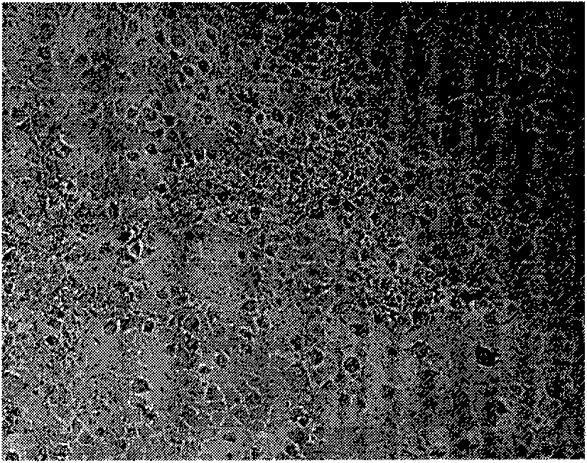
[0055] After transfection, the morphological changes of the cells were observed under a phase-contrast microscope, and compared with primary porcine small intestinal epithelial cells (such as figure 1 )Compare.

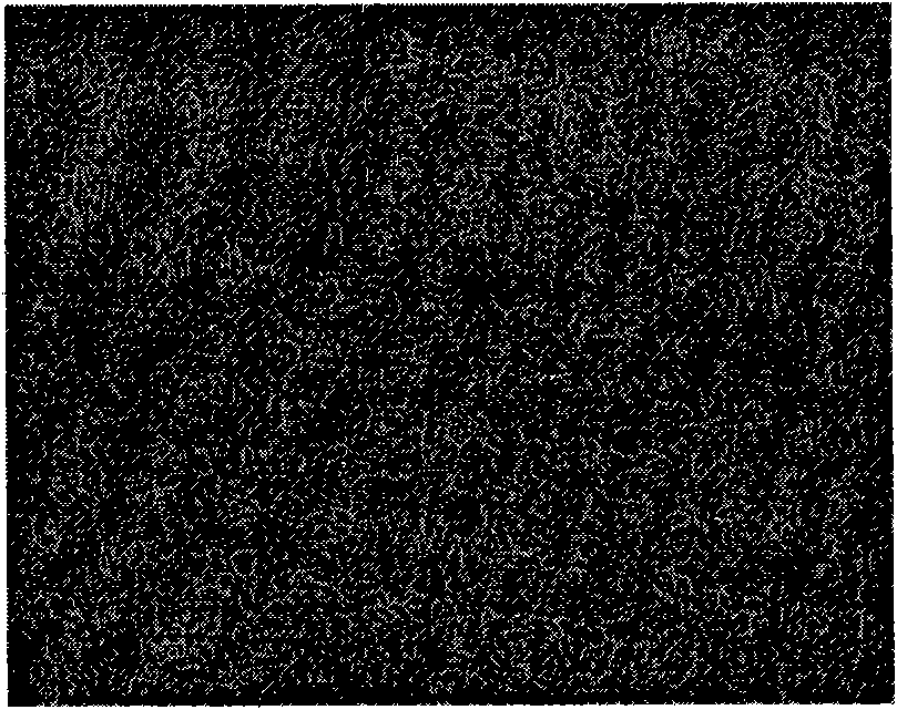
[0056] ZYM-SIEC02 (such as figure 2 ) is similar to the primary cells in shape, the central nucleus of the cells is obvious, and 2 or more nucleoli can be clearly seen. Most of the cells were polygonal or oval in shape, and the cells were arranged in a typical cobblestone-like arrangement, and there was contact inhibition between cells.

Embodiment 3

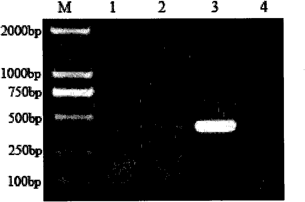
[0057] Example 3.RT-PCR detection of exogenous hTERT expression in transfected cells

[0058] (1) Primer design and synthesis

[0059] Design a pair of specific detection primers according to the human telomerase reverse transcriptase gene reference sequence published in GenBank (Accession No. NM-198255), the upstream and downstream primers are P1: 5′-TATGCCGTGGTCCAGAAG-3′; P2: 5′-TATGCCGTGGTCCAGAAG-3′, the size of the amplified fragment is 416bp, and the primers were synthesized by Beijing Sanbo Polygala Biological Co., Ltd.

[0060] (2) Extraction of total cellular RNA

[0061] ①The transfected cells and the primary cells were digested with ATV and inoculated into 35 mm culture dishes. After the cells covered the monolayer, they were taken out, washed twice with sterilized PBS, and the liquid was discarded.

[0062] ② Add 750 μL Trizol reagent, mix up and down, and after the cells are completely lysed, pipette the cell lysate into a 1.5 mL sterilized EP tube treated with D...


PUM


Abstract

The invention discloses a pig small intestine epithelial cell line (ZYM-SIEC02), which was preserved in China Center for Type Culture Collection on January 13th, 2010 with a collection number of CCTCC-C201001. The invention also discloses the construction method of the cell line. The cell line has the advantages of low nutrient condition requirements, quick cell growth, easy cultivation and convenient promotion and application.

Description

technical field [0001] The invention belongs to the technical field of animal cell engineering and relates to the establishment of a new cell line, in particular to a pig small intestinal epithelial cell line (ZYM-SIEC02) and its establishment method. Background technique [0002] Intestinal epithelial cells (IEC) are the main functional cells of the intestine, which participate in digestion, absorption, immune barrier and stress response of the intestine, and are closely related to the endocrine and exocrine functions of the intestine. Therefore, the isolation and culture of small intestinal epithelial cells is one of the main means to study small intestinal function, nutrient absorption mechanism and its regulation. [0003] Instructor Zhang Yanming, College of Animal Science, Northwest Agriculture and Forestry University, and others successively worked on the isolation and culture method of pig umbilical vein endothelial cells, the preliminary study on the application of ...

Claims


Application Information

Patent Timeline
Patent Type & Authority Applications(China)
IPC IPC(8): C12N5/071C12R1/91
Inventor 张彦明王静刘芳宁郭抗抗
Owner NORTHWEST A & F UNIV
Why Eureka
Try Eureka