
Endoglin antibody coupled liposome as well as preparation method and application thereof

An antibody conjugation and liposome technology, which can be used in liposome delivery, preparations for in vivo tests, and medical preparations with inactive ingredients, etc., and can solve problems such as limited application scope

Inactive Publication Date: 2011-04-13
赵永祥
View PDF4 Cites 17 Cited by
  • Summary
  • Abstract
  • Description
  • Claims
  • Application Information

AI Technical Summary

Problems solved by technology

[0005] However, for the antibodies bound by the immunoliposome materials prepared in the prior art for tumor treatment, the specific antigens only exist in specific types of tumor cells, so this type of liposome materials can only Targeted to specific types of tumor cells, its scope of application is therefore greatly limited

Method used


Image

  • Endoglin antibody coupled liposome as well as preparation method and application thereof
  • Endoglin antibody coupled liposome as well as preparation method and application thereof
  • Endoglin antibody coupled liposome as well as preparation method and application thereof

Examples


Embodiment 1

[0055] Embodiment 1: the preparation particle diameter is the plasmid DNA liposome of Endoglin antibody coupling of 100nm, and specific preparation steps are as follows:

[0056] 1) Mix POPC, DDAB, DSPE-PEG with chloroform 2000 and DSPE-PEG 2000 -Maleimide was formulated into corresponding concentrations of 20mg / mL, 5mg / mL, 20mg / mL and 10mg / mL respectively, and mixed in Agilent chromatographic vials in amounts of 18.6μmol, 0.6μmol, 0.6μmol and 0.2μmol; mix well After that, the organic solvent was removed by nitrogen flow for 15 minutes, and the sample bottle was continuously rotated until a thin layer of phospholipids was formed; then placed in a rotary evaporator for vacuum evaporation for 2 hours.

[0057] 2) Take out the sample bottle, add 0.2mL TRIS-HCL buffer solution (50mM, pH 7.0), rotate and oscillate slightly, place it in a water bath sonicator for 3 minutes, fully resuspend the phospholipid mixture, and try to avoid the generation of air bubbles; Add dropwise enhan...

Embodiment 2

[0069] Embodiment 2: Preparation of liposomes loaded with LSS670 dye (available from Kodak) coupled with Endoglin antibody with a particle size of 100nm, the specific preparation steps are as follows:

[0070] 1) Mix POPC, DDAB, DSPE-PEG with chloroform 2000 and DSPE-PEG 2000 -Maleimide was formulated into corresponding concentrations of 20mg / mL, 5mg / mL, 20mg / mL and 10mg / mL respectively, and mixed in Agilent chromatographic vials in amounts of 18.6μmol, 0.6μmol, 0.6μmol and 0.2μmol; mix well After that, the organic solvent was removed by nitrogen flow for 15 minutes, and the sample bottle was continuously rotated until a thin layer of phospholipids was formed; then placed in a rotary evaporator for vacuum evaporation for 2 hours.

[0071] 2) Take out the sample bottle, add 0.2mL TRIS-HCL buffer solution (50mM, pH 7.0), rotate and oscillate slightly, put it in a water bath sonicator for 3min, fully resuspend the phospholipid mixture, and try to avoid the generation of air bubb...


PUM

PropertyMeasurementUnit
Particle sizeaaaaaaaaaa
Molecular weightaaaaaaaaaa

Abstract

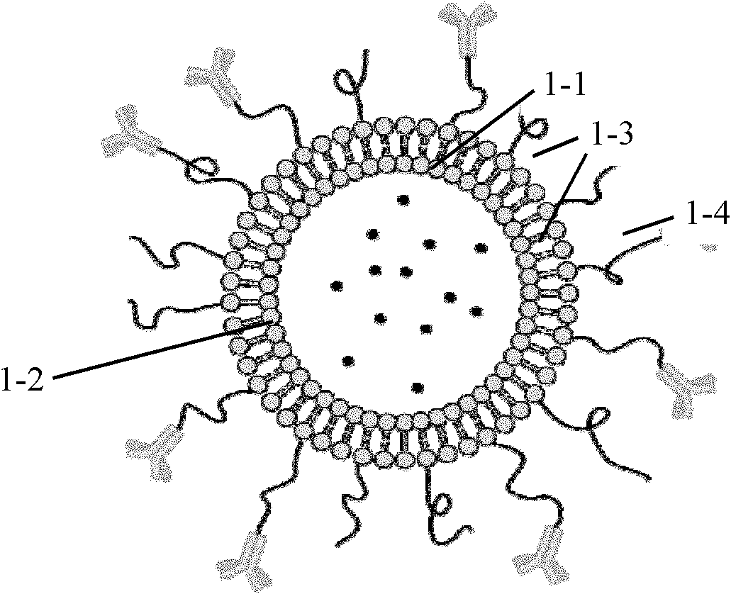
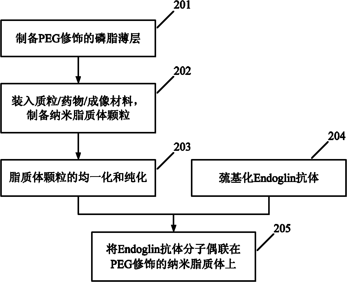
The invention discloses an Endoglin antibody coupled liposome as well as a preparation method and an application thereof, and relates to an Endoglin antibody coupled liposome. The Endoglin antibody coupled liposome comprises nano liposome particles, Endoglin antibody molecules, a coupling agent and a plasmid DNA (deoxyribonucleic acid) / antineoplastic drug / imaging material, wherein the coupling agent is used for coupling the Endoglin antibody onto the nano liposome particles, and the plasmid DNA / antineoplastic drug / imaging material is carried by the nano liposome particles. The preparation method comprises the following steps: preparing a PEG (polyethylene glycol)-modified phospholipid lamina, filling the plasmid / drug / imaging material to prepare the nano liposome particles, homogenizing and purifying the nano liposome particles, thiolating the Endoglin antibody, and coupling the Endoglin antibody molecules on the PEG-modified nano liposome. The Endoglin antibody coupled liposome can be used for preparing drugs for diagnosing and treating solid tumors.

Description

technical field [0001] The invention relates to an Endoglin antibody-coupled liposome, in particular to an Endoglin antibody-coupled liposome and a preparation method and application thereof. Background technique [0002] In tumor diagnosis, drug therapy and gene therapy, finding ideal tumor targets and targeting vectors has always been a research hotspot. [0003] Endoglin is a membrane surface homodimeric glycoprotein molecule (molecular weight of about 180kDa) discovered in the 1980s that is associated with neovascularization of tumors. It is a transforming growth factor-β1 (transforming growth factor-β1, TGF-β1) and cell Accessory molecule for surface receptor binding. After the combination of Endoglin and TGF-β1, it can regulate the downstream signal transduction pathway after the combination of TGF-β1 and its receptor, thereby promoting tumor angiogenesis. Many studies have shown that: Endoglin is mainly distributed in the endothelial cells of new blood vessels and t...

Claims


Application Information

Patent Timeline
IPC IPC(8): A61K9/127A61K47/42A61K48/00A61K49/16A61K49/18A61P35/00A61K47/68A61K47/69
Inventor 赵永祥卓慧钦卢小玲
Owner 赵永祥
Learn More