Singly labeled oligonucleotide fluorescent probe and method for detecting nuclease

A technology of oligonucleotides and fluorescent probes, applied in the field of single-labeled oligonucleotide fluorescent probes and detection of nucleases, can solve the limitations of molecular beacon methods, increase reaction complexity, increase design costs and work to eliminate the disadvantages of unstable hybrids, reduce false positives, and simplify operations

Active Publication Date: 2011-08-17
PEKING UNIV
View PDF0 Cites 28 Cited by
  • Summary
  • Abstract
  • Description
  • Claims
  • Application Information

AI Technical Summary

Problems solved by technology

This not only increases the design cost and workload, but also the additional DNA molecules increase the complexity of the reaction. It is necessary to consider both the concentration matching of the molecular beacon and the complementary template, and the reaction conditions that allow the two to form a stable hybrid.
Many factors limit the practical application of molecular beacon methods in the detection of nucleases

Method used


Image

  • Singly labeled oligonucleotide fluorescent probe and method for detecting nuclease
  • Singly labeled oligonucleotide fluorescent probe and method for detecting nuclease
  • Singly labeled oligonucleotide fluorescent probe and method for detecting nuclease

Examples


Embodiment 1

[0039] Embodiment 1

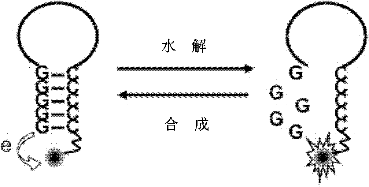
[0040] In this example, detection of exonuclease III (ExoIII) activity was performed using a hydrolysis-mode single-labeled probe. The 5'-end of the single-labeled probe is a continuous cytosine deoxyribonucleotide and labeled with a fluorescent group, and the 3'-end is a continuous guanine deoxyribonucleotide. For the detection principle, see figure 1 In the hydrolysis mode, the specific steps are as follows:

[0041] 1. Hydrolysis mode The initial fluorescent signal of the single-labeled probe is in a quenched state, and it is mixed with Exo III and placed in a suitable solution condition to form a reaction system.

[0042] 2. The 3'-free end (hydroxyl end) of the double-stranded part of the probe is hydrolyzed into mononucleotides base by base by Exo III, and the fluorescent signal is released, which is detected by a real-time fluorescent PCR instrument. As the reaction progresses, the number of probes hydrolyzed increases, and the fluorescence sign...

Embodiment 2

[0051] Example 2

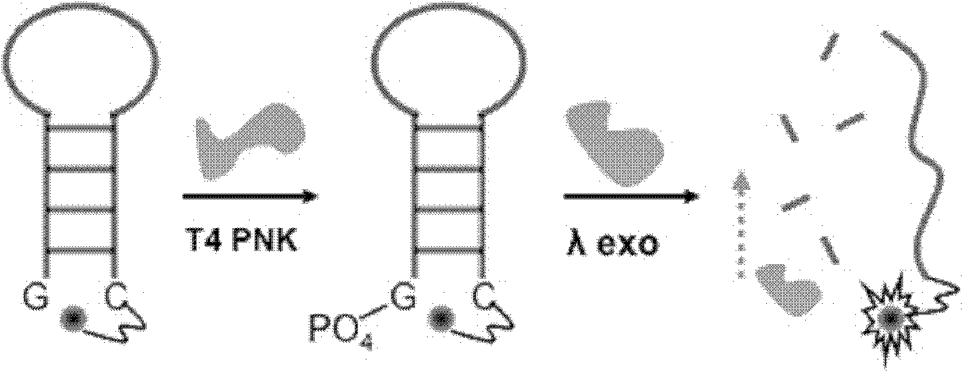
[0052] In this example, detection of T4 polynucleotide kinase phosphorylation activity was performed using a hydrolysis-mode single-labeled probe coupled to a lambda exonuclease. The 5'-end of the single-labeled probe is continuous guanine deoxyribonucleotides with a 5'-hydroxyl terminal, and the 3'-end is continuous cytosine deoxyribonucleotides and labeled with a fluorescent group. For the detection principle, see figure 2 ,Specific steps are as follows:

[0053] 1. The hydrolysis mode single-labeled probe (initially in a state of fluorescence quenching) is mixed with T4 polynucleotide kinase and lambda exonuclease and placed in a suitable solution condition to form a reaction system.

[0054] 2. T4 polynucleotide kinase acts on the stem of the probe to phosphorylate the 5'-terminal base.

[0055] The phosphorylated double-stranded DNA at the 3.5'-terminal base is hydrolyzed by λ exonuclease from the 5'-terminal to a single nucleotide, and the fluoresc...

Embodiment 3

[0067] Embodiment 3

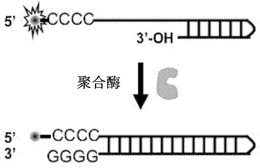
[0068] In this example, detection of Klenow polymerase activity was performed using a synthetic mode single-labeled probe. The 5'-end of the single-labeled probe is a continuous cytosine deoxyribonucleotide side chain and labeled with a fluorescent group. For the detection principle, see image 3 ,Specific steps are as follows:

[0069] 1. Mix the synthetic mode single-labeled probe (the initial fluorescent signal is very strong) with the polymerase and place it in a reaction system with corresponding solution conditions.

[0070] 2. Taking the guanine-quenching fluorescent group generated by the polymerization extension as the indicator signal, the decrease of the fluorescent signal is detected by a real-time fluorescent PCR instrument.

[0071] As the reaction progresses, the number of probes synthesized by extension increases, and the fluorescence signal decreases rapidly until the reaction balances and the fluorescence intensity reaches a plateau v...


PUM


Abstract

The invention discloses a singly labeled oligonucleotide fluorescent probe and a method for detecting nuclease. The singly labeled oligonucleotide fluorescent probe has a stem loop structure, wherein a loop part consists of 5-24 nucleotide residues; a stem part is in a hydrolysis mode or a synthesis mode according to the activity of nuclease to be detected; in the hydrolysis mode, the stem part has a double-chain structure, the tail end is provided with at least three continuous G-C base pairs, the C base ends are labeled with fluorescence groups, the G base ends have quenching effects, one chain of the stem part is hydrolyzed under the action of the nuclease to release a fluorescence signal; and in the synthesis mode, the stem part consists of a section of double chains and a 5'-(dC)4-8side chain, the 5'- end is labeled with a fluorescence group, the 3'- end reacts with the nuclease, 4-8 continuous guanine deoxyribonucleotides are produced by polymerization extension, the fluorescence group at the 5'- end is quenched, and the activity of the nuclease to be detected is analyzed according to the change condition of the fluorescence signal.

Description

technical field [0001] The present invention relates to the field of analysis and detection of various nucleases such as exonuclease, polymerase, polynucleotide kinase, phosphatase, more specifically, relates to a single-labeled oligonucleotide fluorescent probe, and the use of A single-labeled oligonucleotide fluorescent probe assay for the detection of nucleases. Background technique [0002] DNA is an important genetic material for the transmission of life information, and this transmission process depends on the special functions of various nucleases. For example, important life processes such as nucleic acid extension, correction, ligation, phosphorylation, dephosphorylation, and enzyme cleavage all rely on the specific interaction between nucleases and DNA. A quick and easy nuclease kinetic analysis method is of great significance for the study of biodiversity and the in-depth understanding of life phenomena. [0003] For the analysis of nuclease activity, there are ...

Claims


Application Information

Patent Timeline
Patent Type & Authority Applications(China)
IPC IPC(8): C12Q1/68
Inventor 赵美萍宋晨张晨苏昕
Owner PEKING UNIV
Why Eureka
Try Eureka