

Molecular markers of well-differentiated early liver cancer and use thereof
A molecular marker, early-stage liver cancer technology, applied in the field of molecular markers of well-differentiated early-stage liver cancer, can solve the problem of no difference in the expression of SQSTM1, and achieve the effect of high detection sensitivity and specificity
- Summary
- Abstract
- Description
- Claims
- Application Information
AI Technical Summary
Problems solved by technology
Method used
Image

Examples
Embodiment 1
[0016] Example 1: Collection of samples and protein extraction:
[0017] 1. Human atypical hyperplasia nodular lesions and early liver cancer lesions were diagnosed under the microscope by HE slices of 2 pathologists, and the corresponding wax blocks were cut into white slices with a thickness of 7 μm. Dry at 60°C until there are no air bubbles between the sliced tissue and the glass slide (about 30-60 minutes).
[0018] 2. Formaldehyde-fixed / paraffin-embedded (FFPE) tissue protein extraction and protein concentration determination:
[0019] 2.1. Dewax the above-mentioned liver tissue sections in xylene for 2 times, 10 min each time. After air-drying, the range of the lesion is accurately drawn. Use a scalpel blade (No. 11 blade) to cut the lesion precisely and put it into a low adsorption tube.
[0020] 2.2. Add an appropriate amount of paraffin-embedded tissue protein extraction buffer (Tris-HCl buffer) to each low adsorption tube, which contains 2% sodium dodecylsulfon...
Embodiment 2
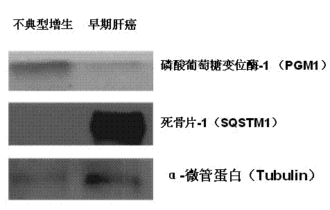
[0023] Example 2: Immunoblot method proves the expression difference of phosphoglucomutase-1 and sequestrum-1:
[0024] 1. The protein samples extracted in Example 1 (mixed with 18 samples in each group) were subjected to sodium dodecylsulfonate-polyacrylamide gel electrophoresis (SDS-PAGE) by conventional methods, and then transferred to PVDF membrane (Millipore) on.
[0025]2. Dissolve skim milk powder in TBS-T buffer (0.05% (v / v) Tween 20 / Tris-buffer) at pH=7.4 to obtain fat-free milk solution, the concentration of skim milk powder is 5% (w / v ), add the above fat-free milk solution to the above protein sample, add the primary antibody after 1h, incubate overnight at 4°C, wash the membrane with TBS-T for 3 times, add horseradish peroxidase (HRP)-labeled secondary Antibody, incubated at room temperature for 1h. Among them, the company and dilution ratio of the primary antibody are as follows: mouse anti-PGM1 (ab55616, Abcam, 1:300), rabbit anti-SQSTM1 (P0067, Sigma-Aldrich,...
Embodiment 3
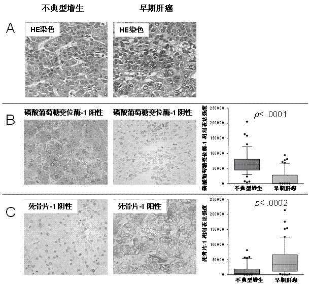
[0027] Example 3: Immunohistochemical method proves the expression difference of PGM1 and SQSTM1:
[0028] 1. After taking human atypical hyperplasia nodular lesions and early liver cancer lesions and diagnosing them under the microscope with the HE slices of two pathologists, the corresponding wax blocks were made into tissue chips by conventional methods, cut into 4 μm thick slices, and placed onto 3-aminopropyltriethoxysilane-coated glass slides.
[0029] 2. Dewax the slides with tissue sections according to conventional methods, and then hydrate them with 100% ethanol, 95% ethanol, and 85% ethanol for 5 minutes each. Place the slides with tissue sections in In 0.01M citrate buffer (pH6.0), carry out antigen retrieval treatment at 100°C for 3 minutes, take it out and let it cool down, and place it in H2O with a mass concentration of 3%. 2 o 2 Endogenous peroxidase was blocked in phosphate (PBS) buffer solution for 30 minutes, removed, placed in goat serum for 60 minutes, ...
PUM
| Property | Measurement | Unit |
|---|---|---|
| Thickness | aaaaa | aaaaa |
Abstract
Description
Claims
Application Information