Bionic bone repair scaffold body with layered structure and preparation method
A layered structure and scaffold technology, applied in prostheses, tissue regeneration, bone implants, etc., can solve problems such as difficulty in ensuring the mechanical properties of scaffolds, damage to scaffold materials, and unsuitability for clinical applications, etc., to achieve flexible adjustment of three-dimensional Dimensions, effect of mechanical transmission, good structural stability
- Summary
- Abstract
- Description
- Claims
- Application Information
AI Technical Summary
Problems solved by technology
Method used
Image
Examples
Embodiment 1
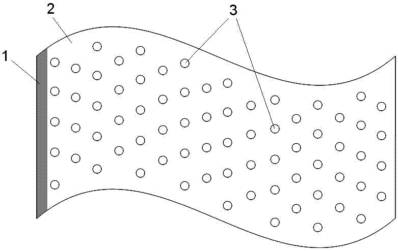
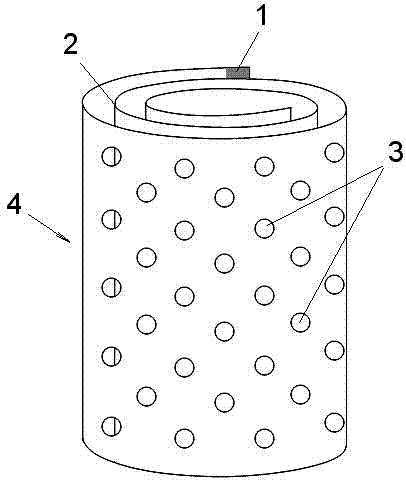
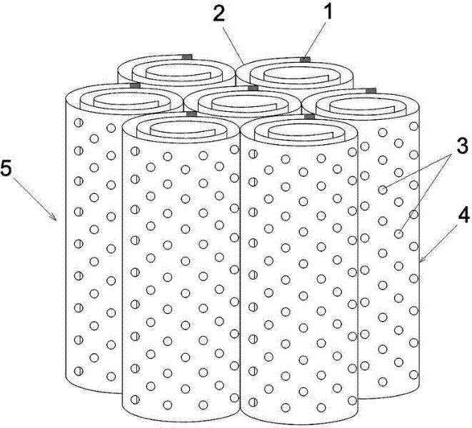
[0045] Dissolve 2g of chitosan in 50mL of acetic acid solution with a concentration of 2 (v)%, and stir fully to completely dissolve and evenly disperse chitosan. Add 1g Tween-20 and 1g Tween-80 to the chitosan solution, stir continuously at 1000 rpm for 30 minutes, quickly pour the foam-rich film-forming solution into a glass plate, and spread it horizontally to form a film In flake form, freeze at -20°C and dry in vacuum. Soak with 1 (w)% NaOH solution to neutralize the residual acetic acid in the membrane, fully wash with distilled water and freeze-dry to obtain a porous chitosan membrane material 2 containing pores 3, such as figure 1 As shown, the thickness in the wet state is 0.40mm. The membrane material 2 is fully water-absorbed and swollen, cut into a suitable size, and tightly spirally wound, and the edge part 1 of the end wrapping is eroded with a small amount of 2 (v)% acetic acid solution to dissolve the surface, and glued to the spirally wound cylinder Body-sha...
Embodiment 2
[0047] Dissolve 2g of polycaprolactone in 50mL of dimethyl sulfoxide, add 50g of 40-60 mesh NaCl particles, stir well to make the salt particles evenly distributed, and make a viscous film-forming liquid. Pour the film-forming solution into a glass plate, spread it horizontally into a film, and dry it in vacuum at 50°C. Soak and wash with distilled water repeatedly to remove NaCl particles in the membrane to obtain a porous polycaprolactone membrane material 2 with a thickness of 0.21 mm in a wet state. The membrane is cut to a suitable size, tightly spirally wrapped, and the end edge part 1 is eroded with a small amount of dimethyl sulfoxide on the surface, and bonded to the spirally wrapped cylinder. After drying, thoroughly soak and rinse with distilled water to remove residual solvents. A polycaprolactone porous scaffold unit 4 with a helically wound cylindrical structure was obtained, the pores 3 interconnected with each other, the average pore diameter was 223.1 μm, and...
Embodiment 3
[0049] Under the catalysis of calcium chloride, dissolve 10g of polyamide 66 in 100mL of absolute ethanol solution at 70°C to 80°C, and stir well to make a viscous film-forming liquid. After cooling to room temperature, the film-forming solution was poured into a glass plate, spread horizontally into a film, and dried at 60°C with a thickness of 0.15 mm. Repeated soaking and washing with distilled water, and drying at 70° C. to obtain porous polyamide 66 membrane material 2 . The membrane is cut to a suitable size, tightly spirally wrapped, and the end edge part 1 is etched with a small amount of ethanol solution containing calcium chloride on the surface, and bonded to the spirally wrapped cylinder. After drying, thoroughly soak and rinse with distilled water to remove residual solvents. A polyamide 66 porous scaffold unit 4 with a helically wound cylindrical structure was obtained, the pores 3 interconnected with each other, the average pore diameter was 68.0 μm, and the po...
PUM
| Property | Measurement | Unit |
|---|---|---|
| diameter | aaaaa | aaaaa |
| thickness | aaaaa | aaaaa |
| pore size | aaaaa | aaaaa |
Abstract
Description
Claims
Application Information
 - R&D
- Intellectual Property
- Life Sciences
- Materials
- Tech Scout
- Unparalleled Data Quality
- Higher Quality Content
- 60% Fewer Hallucinations
Browse by: Latest US Patents, China's latest patents, Technical Efficacy Thesaurus, Application Domain, Technology Topic, Popular Technical Reports.
© 2025 PatSnap. All rights reserved.Legal|Privacy policy|Modern Slavery Act Transparency Statement|Sitemap|About US| Contact US: help@patsnap.com


