Intracellular protein detection method by flow cytometry

A flow cytometry, intracellular protein technology, applied in biological testing, material testing, etc., to achieve the effect of strong surface adsorption, simple preparation and good stability

Active Publication Date: 2012-06-27
JILIN UNIV
View PDF4 Cites 4 Cited by
  • Summary
  • Abstract
  • Description
  • Claims
  • Application Information

AI Technical Summary

Problems solved by technology

[0005] In recent years, researchers have discovered that quantum dot nanoparticles have bioluminescent properties, which can be used as labeling carriers to detect tumors by flow cytometry, but this method can only detect specific molecules on the cell surface

Method used


Image

  • Intracellular protein detection method by flow cytometry
  • Intracellular protein detection method by flow cytometry

Examples


Embodiment 1

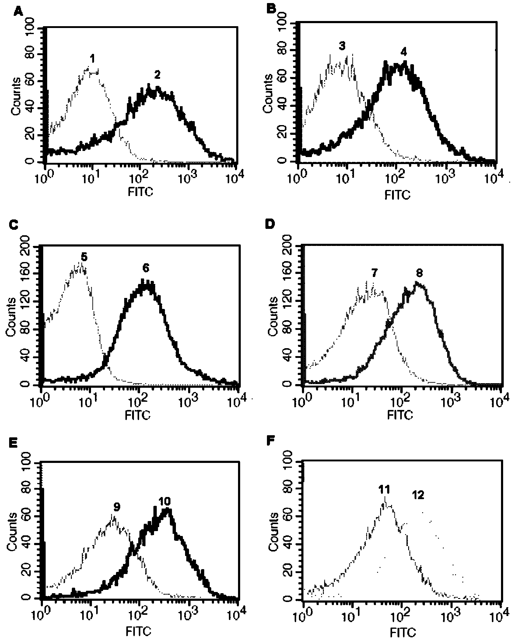
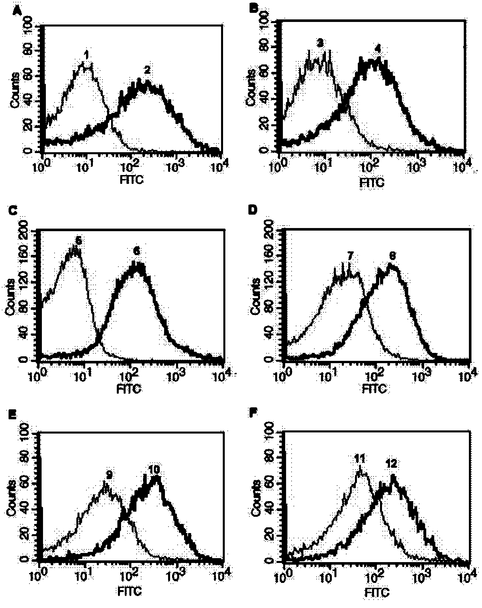
[0040] Example 1: Take the expression of actin (β-actin) in the human liver cancer HepG2 cell line as an example to illustrate.

[0041] 1. Preparation of Silica Nanoparticles

[0042] (1) Preparation of surface aminated silica nanoparticles:

[0043] adopt first Law( W, Fink A, Bohn E.J Colloid Interface Sci, 1968, 26~62.) prepare 100~120nm silica nanoparticles; then add about 1g of silica nanoparticles to 30ml of anhydrous toluene, stir at room temperature, and then Slowly add 2.5ml of γ-aminopropyltriethoxysilane (ATES) dropwise, continue to stir for 1h, then raise the temperature to 95°C, reflux for 10h, wash with isopropanol ultrasonically for 3 times, and vacuum dry at 60°C for 10h to obtain about 1g surface aminated silica nanoparticles;

[0044] (2) Preparation of surface carboxylated silica nanoparticles:

[0045] adopt first Law( W, Fink A, Bohn E.J Colloid Interface Sci, 1968, 26~62.) Prepare 100~120nm silica nanoparticles; then uniformly disperse 0.01M s...

Embodiment 2

[0056] Example 2: Take the expression of protein kinase B (Akt2) in human breast cancer MCF-7 cell line as an example to illustrate.

[0057] 1. the preparation of silicon dioxide nanoparticle (same as embodiment 1)

[0058] 2. Preparation of nanoparticles with specificity for binding intracellular proteins

[0059] (1) Preparation of surface-aminated silica nanoparticles with specificity for binding intracellular proteins: first suspend the surface-aminated silica nanoparticles of 100-120 nm in a glutaraldehyde solution with a mass fraction of 25%, Make the final concentration 1mg / ml, stir overnight at room temperature to activate the particles; wash 3 times with 3ml PBS (pH 8.0) the next day to remove excess glutaraldehyde solution; then resuspend in PBS (pH 8.0) to form 2.5mg / ml nanoparticle suspension;

[0060] Take 100 μl of anti-Akt2 antibody solution (purchased from Santa Cruz Biotechnology, derived from mice, antibody:PBS volume ratio = 1:800) and add it to the above...

Embodiment 3

[0068] Example 3: The expression of phosphorylated protein kinase B (p-Akt1 / 2 / 3) in the human cervical cancer Hela cell line is illustrated as an example.

[0069] 1. the preparation of silicon dioxide nanoparticle (same as embodiment 1)

[0070] 2. Preparation of nanoparticles with specificity for binding intracellular proteins

[0071] (1) Preparation of surface-aminated silica nanoparticles with specificity for binding intracellular proteins: first suspend the surface-aminated silica nanoparticles of 100-120 nm in a glutaraldehyde solution with a mass fraction of 25%, Make the final concentration 1mg / ml, stir overnight at room temperature to activate the particles; wash 3 times with 3ml PBS (pH 8.0) the next day to remove excess glutaraldehyde solution; then resuspend in PBS (pH 8.0) to form 2.5mg / ml nanoparticle suspension;

[0072] Add 100 μl of anti-p-Akt1 / 2 / 3 antibody solution (purchased from Santa Cruz Biotechnology, derived from mice, antibody: PBS volume ratio = 1:...


PUM

PropertyMeasurementUnit
particle diameteraaaaaaaaaa
concentrationaaaaaaaaaa

Abstract

The invention belongs to the field of flow cytometry technology, and specifically relates to an intracellular protein detection method by the use of flow cytometry. The method comprises the following steps of: connecting an amino or carboxy functional group on the surface of 100-120nm silica nanoparticle with an amino group of an antibody against the intracellular protein to be detected by the use of covalent bonds, incubating the silica nanoparticle together with intracellular proteins obtained by ultrasonic and other methods, adding another differently sourced antibody which targeted the same intracellular protein to be detected and carrying out incubating, then incubating with a fluorescein labeled second antibody to form a silica nanoparticle-antibody-intracellular protein-antibody-fluorescein labeled second antibody compound, and finally carrying out qualitative detection on the compound by using flow cytometry. According to the invention, after binding to the antibody against the intracellular protein to be detected, the silica nanoparticle has initiative targeting ability and can target various different intracellular proteins.

Description

technical field [0001] The invention belongs to the technical field of flow cytometry, and in particular relates to a method for detecting intracellular proteins by using flow cytometry. Background technique [0002] Malignant tumor, also known as cancer, is one of the major diseases threatening human life and health. The incidence of cancer is increasing year by year, especially after the 1970s, the number of cancer incidence is increasing at an average annual rate of 3% to 5%. Not only that, the age of onset of cancer is also tending to be younger, and it has become the second leading cause of death in humans . For this reason, the medical profession has been working hard to find an effective diagnosis and treatment for it. After more than half a century of development and improvement, surgery, radiotherapy, chemotherapy and other methods have become the main means of comprehensive treatment of malignant tumors, but they often cause serious side effects in clinical appli...

Claims


Application Information

Patent Timeline
Patent Type & Authority Applications(China)
IPC IPC(8): G01N33/68
Inventor 施维于洋朱广山李全顺曹秉振
Owner JILIN UNIV
Why Eureka
Try Eureka