Device for fixing experimental animal during perfusion and application thereof

A technology for fixing devices and experimental animals, applied in the direction of restraining animals, veterinary instruments, medical science, etc., can solve the problems of human harm, explosion, non-compliance with laboratory safety protection standards, etc., to ensure the perfusion effect and protect health. Effect

Inactive Publication Date: 2014-12-17
PEOPLES HOSPITAL OF XINJIANG UYGUR AUTONOMOUS REGION
View PDF6 Cites 0 Cited by
  • Summary
  • Abstract
  • Description
  • Claims
  • Application Information

AI Technical Summary

Problems solved by technology

[0004] (1) The simple perfusion device cannot provide the necessary protection for the experimenter: the perfusion fixative used in the perfusion fixation method is mostly formaldehyde or paraformaldehyde, and the experimenter is directly exposed to formaldehyde vapor during operation, which will cause serious harm to the human body and does not meet the requirements. Laboratory safety protection specification requirements
[0005] (2) Manual control of the syringe is not easy to control the pressure: when the perfusion fixation method uses a simple device combined with a syringe and injection needle, the pressure is manually controlled by the operator. If the pressure is too small: the perfusion effect is not good; if the pressure is too large: it will cause tissue damage (eg, burst blood vessel, burst heart)
When using a simple device combined with an infusion bottle and an injection needle, it is suitable for rats and rabbits, but the pressure of the device is significantly lower in mice or gerbils due to the small blood vessels, and the perfusion effect cannot be ensured.

Method used


Image

  • Device for fixing experimental animal during perfusion and application thereof

Examples


Embodiment 1

[0020] Embodiment 1: Experimental animal perfusion fixation device and its application

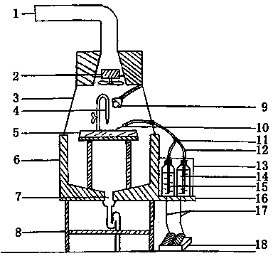
[0021] See attached figure 1 As shown, a perfusion fixture for experimental animals includes an exhaust pipe (1), an exhaust fan (2), a plexiglass cover (3), a test nozzle (4), a perfusion table (5), a water tank (6 ), drainage hole (7), bracket (8), spotlight (9), perfusion needle (10), tee tube (11), infusion hose (12), liquid pump (13), saline tank (14) , a fixed liquid tank (15), a liquid pump placement platform (16), a liquid pump control pedal cable (17), and a liquid pump control pedal (18); among them, the bracket (8) supports the water tank (6) and the right side set The liquid pump placement platform (16), the water tank (6) and the liquid pump placement platform (16) are integrated with the same bottom surface, and the bottom of the water tank (6) is inclined at 5 degrees to the center drain hole (7). A perfusion table (5) is installed inside, and the front of the table is i...

Embodiment 2

[0028] Example 2: Application of perfusion fixation device in the study of focal lymphatic stagnant encephalopathy model in rats

[0029]After the male SD rats were anesthetized, the top of the skull was prepared and disinfected, and the scalp was incised 2 cm longitudinally in the middle, and the periosteum was corroded by 30% hydrogen peroxide to expose the bregma. On the surface of the meninges, uncover the dura mater, expose the arteries on the surface of the brain, use 3 / 8 needled medical nylon thread to separate and prepare the thread, half-ligate the artery, suture the scalp after the operation, establish the animal model of focal lymphatic stagnant encephalopathy, and use it 24 hours after the operation The perfusion fixation device performs transcardiac perfusion brain extraction. First, put normal saline and 4% paraformaldehyde into the normal saline tank (14) and the fixed liquid tank (15); turn on the power supply of the exhaust fan (2) and the spotlight (9); adj...

Embodiment 3

[0030] Example 3: Application of perfusion fixation device in research on ultrastructure of lung tissue in guinea pig model of asthma

[0031] Male guinea pigs (250-500 g) were sensitized by intraperitoneal injection of 100 mg / kg oval protein solution at a mass concentration of 1 kg / L, and 3 weeks later were atomized and inhaled with a mass concentration of 0.1 kg / L oval protein solution to establish asthmatic guinea pigs When guinea pigs have symptoms such as shortness of breath, lip cyanosis, dyspnea and even convulsions, the perfusion fixation device can be used to obtain materials through cardiac perfusion. Normal saline tank (14) and fixed fluid tank (15); turn on the exhaust fan (2) and spotlight (9) power supply; adjust the pressure of the equipment, lightly step on the liquid pump control pedal (18), pump out normal saline, and make the infusion The hose (12) and the perfusion needle (10) are filled with liquid and the air is drained; place the guinea pig on the perf...


PUM


Abstract

The invention discloses a device for fixing an experimental animal during perfusion and an application thereof. A bracket is used for supporting a water groove and a liquid pump accommodating stage arranged on a right side, wherein the water groove and the liquid pump accommodating stage share the same bottom surface, and are integrated; the periphery of the bottom of the water groove inclines towards a drainage hole in the center; a perfusion stage is arranged in the water groove; the front side of the stage surface of the perfusion stage inclines towards an operator; an assay water nozzle and a light collecting lamp are arranged above the perfusion stage; an organic glass cover and an exhaust fan are arranged above the water groove; the organic glass cover is used for fixing the exhaust cover; the organic glass cover is connected with an exhaust pipe; the liquid pump accommodating stage is used for accommodating a liquid pump; the liquid pump is connected with a liquid pump control pedal through a liquid pump control pedal cable; a transfusion hose is inserted into two liquid supply units, i.e., a normal saline solution water tank and a fixed liquid tank respectively; two liquid supply units are arranged in the liquid pump; and the transfusion hose is connected with a perfusion needle through a three-way pipe. The device has the characteristics of simple structure, convenience for using, safety, operability by one person, high working efficiency and the like, and has wide application value.

Description

technical field [0001] The invention relates to the technical field of an experimental animal perfusion device, in particular to the technical field of an experimental animal perfusion fixation device and its application. Background technique [0002] In the histopathological research of experimental animals, especially in situ hybridization and immunohistochemical research, in order to avoid or reduce the changes in tissue morphology and structure after death, the perfusion fixation method is often used before the animals are sacrificed. Good fixation can prevent the autolysis and corruption of tissue cells, and keep the protein, fat, sugar, enzyme and other components in the cells similar to those in life; with the perfusion fixation method, the fixative can be injected into the blood vessels in time, and reach the whole body through the blood vessel branches. Tissue, so that the tissue is fully fixed, so in the study of histomolecular pathology, perfusion fixation is most...

Claims


Application Information

Patent Timeline
Patent Type & Authority Patents(China)
IPC IPC(8): A61D7/00A61D3/00
Inventor 补娟朱沂史深莎丽娅那色尔李红燕杨雪荣张拥军党辉陈浩吴波
Owner PEOPLES HOSPITAL OF XINJIANG UYGUR AUTONOMOUS REGION
Why Eureka
Try Eureka