Substrate with photo-controllable cell adhesion property, method for analyzing and fractionating cells, and device for analysis and fractionation of cells

An adhesive material and adhesive technology, applied in the field of stem cell research and regenerative medicine, can solve the problems of reduced survival rate, fluorescence correction, complex optical axis adjustment, cell damage, etc., and achieve the effect of improving purity and simple real-time operation.

Inactive Publication Date: 2012-09-12
HITACHI HIGH-TECH CORP
View PDF7 Cites 7 Cited by
  • Summary
  • Abstract
  • Description
  • Claims
  • Application Information

AI Technical Summary

Problems solved by technology

Multiple fluorescent markers can be resolved by irradiating multi-colored lasers, but fluorescence correction and optical axis adjustment are complicated
In addition, if trypsin treatment is performed to separate the cell mass into individual cells in advance, it will cause considerable damage to the cells.
Furthermore, although the sorted cells have high purity and recovery rate, there is a problem that the survival rate decreases due to the shock during sorting.

Method used


Image

  • Substrate with photo-controllable cell adhesion property, method for analyzing and fractionating cells, and device for analysis and fractionation of cells
  • Substrate with photo-controllable cell adhesion property, method for analyzing and fractionating cells, and device for analysis and fractionation of cells
  • Substrate with photo-controllable cell adhesion property, method for analyzing and fractionating cells, and device for analysis and fractionation of cells

Examples


Embodiment 1

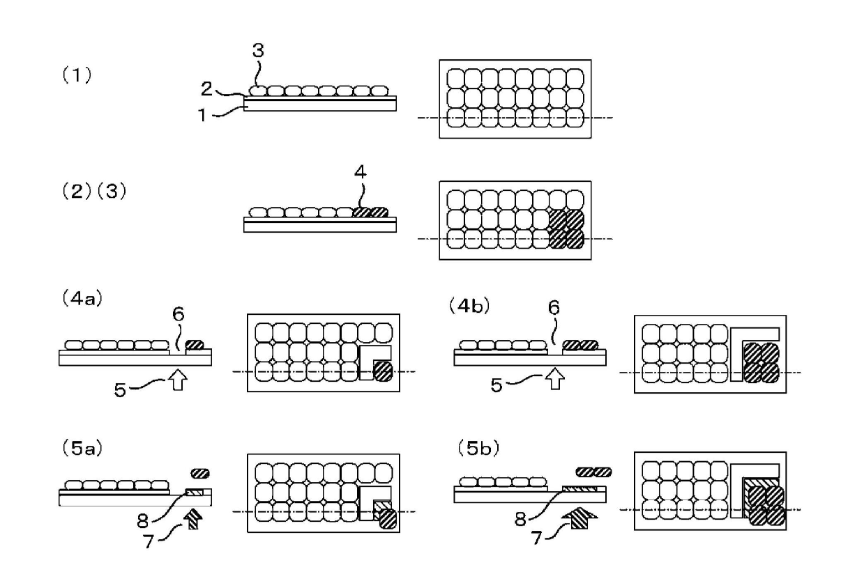
[0112] Prepare the methacrylic polymer represented by the general formula (1) (R 1 :Methyl group, n:1) 20 mol%, methacrylic polymer represented by general formula (2) (R 1 : Methyl, R 2 : Butylene) 50 mol% and the methacrylic polymer represented by the general formula (10) (R 1 : Methyl, R 6 : OCOCH 2 OCH 2 CH 2 OCH 2 CH 2 O, R 4 : Br, X: CH 2 CO 2 H, n: 1) 30 mol% terpolymer (molecular weight: 5,000 to 50,000) membrane glass culture vessel. Add a cell suspension of human bone marrow stromal cells and human adipocyte differentiation medium (Cell Applications) to it, 2 Culture in an incubator. At the stage when it became 40% full, the glass culture container was installed in the apparatus of the present invention, and the light of 450 nm or less was blocked for microscopic observation. Use the monitor to confirm the position of the cells considered to be abnormal, and set the periphery of the abnormal cell group as the laser ablation area (refer to figure 1 (1)(2)(3)(4a)(5a)). P...

Embodiment 2

[0115] After continuing the culture of the sample of Example 1, the glass culture container was taken out of the incubator. The cells were washed with PBS and treated with a blocking solution for cell surface labeling (JRH) for 1 hour. In order to detect the mesenchymal stem cell marker: CD105, a cell surface labeling blocking solution dilution solution of mouse anti-human CD105 antibody (abcam) was added, and the reaction was carried out at room temperature for 1 hour. After washing with PBS, the cell surface labeling blocking solution diluent of Alexa Fluor 488 labeled anti-mouse IgG antibody (Invitrogen) was added, and the reaction was light-shielded for 1 hour. After the reaction, the solution was replaced with PBS. Next, in order to detect fat cells, OilRed O staining solution (Sigma) was added and left at room temperature for 1 hour for staining, and the solution was replaced with PBS. The observation and differentiation of the cells are performed as follows. A glass c...

Embodiment 3

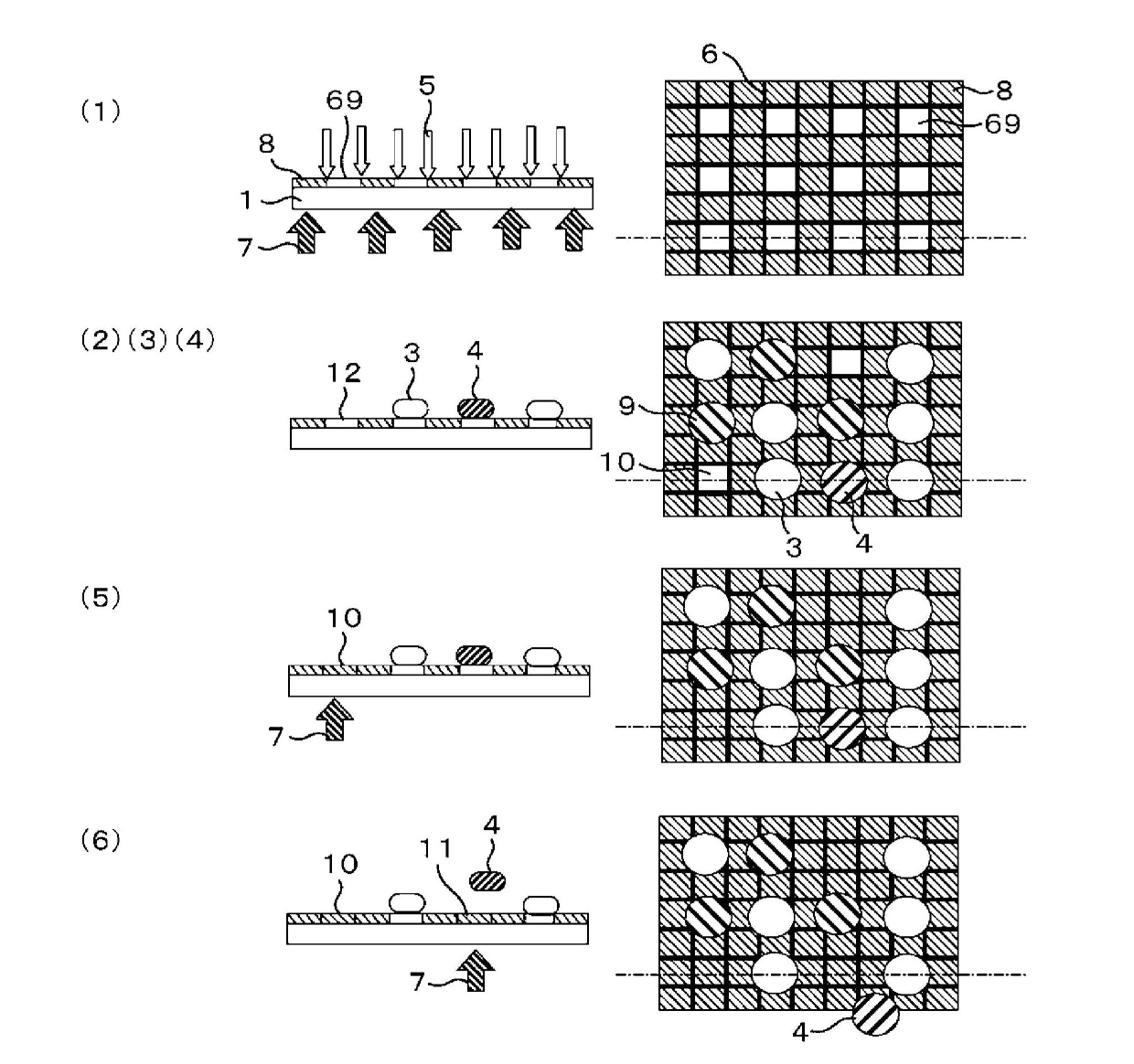
[0118] HBSS was added to another glass culture container cultured in the same manner as in Example 2 and washed, and a Trypsin / EDTA solution was added, and it was left at room temperature for several minutes to peel the cells from the glass culture container. Then, the tripsin neutralization solution was added to stop the reaction, the cells were recovered into a centrifuge tube by flushing, and centrifuged for a few minutes, the supernatant was removed, and the culture medium was added to make a cell suspension. In addition, the application of mesenchymal stem cell markers and adipocyte staining were also performed in the same manner as in Example 2. Next, prepare an alkoxysilane represented by general formula (11) (R 2 : (CH 2 CH 2 O) 2 CH 2 CH 2 , R 3 : Methyl, R 4 : Br, R 5 : O, R 6 : OCOCH 2 O(CH 2 CH 2 O) 3 , X: CH 2 NH 2 ) A membrane glass culture vessel with PBS added and set in the device of the present invention. Then, the cell adhesion areas of 70 μm square are arran...


PUM


Abstract

When cells are analyzed, fractionated, and incubated while keeping the cells alive, real-time operations can be performed more easily and the cells can be incubated while removing unnecessary cells from the incubated cells to purify the cells being incubated. Furthermore, desired cells are separated through analysis from the incubated cells, and the purity, recovery, and viability of the cells are heightened. Use is made of a substrate having photo-controllable cell adhesion properties, the substrate comprising a transparent base and, formed thereon, a film of a material which has photo-controllable cell adhesion properties and has been obtained by bonding a cell-adhesive material to a cell-non-adhesive material through photo-dissociable groups. Cell images are detected and analyzed to obtain information about the location of desired cells. On the basis of the information, a space is formed between cells and the material having photo-controllable cell adhesion properties is cut, by means of second light irradiation. Meanwhile, by means of first light irradiation, the surface of the substrate is changed from a cell-adhesive surface to a cell-non-adhesive surface, thereby separating the cell(s) from the substrate. Thus, cells can be analyzed and fractionated while keeping the cells alive.

Description

Technical field [0001] The invention relates to the field of regenerative medicine and stem cell research, in particular to cell analysis and differentiation and culture technology. Background technique [0002] In the field of regenerative medicine, the following operations are carried out: identification and isolation of very few adult stem cells and precursor cells contained in somatic cells, and then culture them to produce somatic cells. In addition, attempts have been made to culture into adult stem cells or somatic cells by inducing differentiation from iPS cells and ES cells that are sources of adult stem cells. However, these iPS cells and ES cells are not uniform, and the cells induced by their differentiation are not uniform. Produce various cells. Cells or tissues for regenerative medicine are required to be homogeneous as somatic cells, including adult stem cells, and not containing pluripotent stem cells such as cancer cells, cancer stem cells, iPS cells, and ES c...

Claims


Application Information

Patent Timeline
Patent Type & Authority Applications(China)
IPC IPC(8): C12N11/00C12M1/34C12M1/42C12N11/08C12N13/00C12Q1/02G01N33/48
CPCC12M47/04C12N1/02C12N11/02
Inventor 杉山寿高桥智内田宪孝小泽理
Owner HITACHI HIGH-TECH CORP
Why Eureka
Try Eureka