Preparation method for dendritic cell of umbilical cord blood source and dendritic cell vaccine

A technology of dendritic cells and umbilical cord blood is applied in the preparation of DC vaccines and in the field of tumor vaccine treatment products.

Active Publication Date: 2012-09-19
北京和泽普瑞生物科技有限公司 +1
View PDF3 Cites 14 Cited by
  • Summary
  • Abstract
  • Description
  • Claims
  • Application Information

AI Technical Summary

Problems solved by technology

[0004] The present invention solves the problems of low number of DCs derived from peripheral blood and poor antigen presentation ability by improving the selection of cell sources, optimization of culture schemes, extraction and selection of tumor-specific antigens, etc.

Method used


Image

  • Preparation method for dendritic cell of umbilical cord blood source and dendritic cell vaccine
  • Preparation method for dendritic cell of umbilical cord blood source and dendritic cell vaccine
  • Preparation method for dendritic cell of umbilical cord blood source and dendritic cell vaccine

Examples


Embodiment 1

[0017] Example 1 Preparation of DCs derived from umbilical cord blood

[0018] Preparation of autologous cord blood plasma:

[0019] 1. Aseptically collect a portion of umbilical cord blood from a full-term normal delivery fetus after umbilical cord clamping, anticoagulate with citric acid, and centrifuge in a centrifuge tube for 15 minutes.

[0020] 2. Take about 30mL of the supernatant, put it into a centrifuge tube, continue to centrifuge for 15 minutes, and collect the supernatant plasma.

[0021] 3. Place the collected plasma in a 56°C water bath for 30 minutes to inactivate complement.

[0022] 4. Centrifuge to remove the flocculent sediment in the tube, transfer the supernatant to a new centrifuge tube, aliquot and freeze for later use.

[0023] Isolation of cord blood mononuclear cells:

[0024] 1. Mix the umbilical cord blood separated from the upper layer of plasma with normal saline at a ratio of 1:1, and then mix with 6.0% (w / v) hydroxyethyl starch (HESpan) at...

Embodiment 2

[0036] Example 2 Immunophenotypic detection of cultured cells

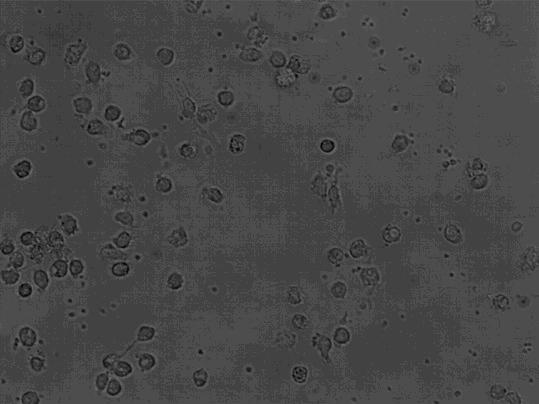
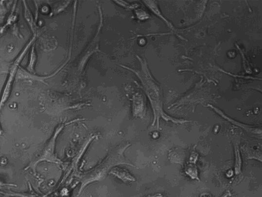
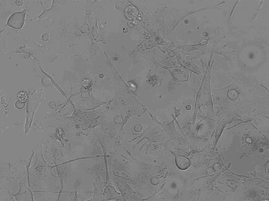
[0037] Take the cells on the 1st, 7th, and 9th day of culture respectively, wash them twice with calcium and magnesium-free PBS, and take 1×10 5 / mL, respectively added to the corresponding FCM tube. Add 5 μl of monoclonal antibodies to be detected, including CD1α, HLA-DR, CD80, CD83, and CD86 antibodies, incubate at 4 °C in the dark for 30 minutes, shake once every 10 minutes, so that the cells can fully contact with the antibodies. Washed twice with PBS, resuspended in 400 μl of PBS, and detected by flow cytometer FASCSCalibur (BD Biosciences), the results are shown in Figure 2, Table 1, image 3 .

[0038]

[0039] Table 1 Immunophenotype of DCs at different culture times

[0040] Training time Day 1 day 7 Day 9 CD1α+ 0.84±0.566% 23.33±2.17% 13.46±1.97% CD83+ 27.37±2.11% 38.57±1.99% 40.88±2.60% HLA-DR+ 76.21±3.09% 82.63±1.62% 89.01±2.59% CD80+ 8.01±2.01% 46....

Embodiment 3

[0042] Example 3 Mixed Lymphocyte Reaction (MLR)

[0043] 1. Take the DCs cultured on day 9, suspend them with AIM-V lymphocyte medium, and adjust the cell concentration to 1×10 6 cells / ml, first treated with mitomycin 25 μg / ml for 45 minutes, and washed with PBS more than 3 times.

[0044] 2. Adjust the DC density to 1×10 5 cells / ml, mixed with T lymphocytes of the corresponding culture time at the ratio of (DC: lymphocytes) 1:10, 1:20, 1:50, 1:100, no DC was added to negative wells, and 3 cells were set in each group Duplicate wells and culture for 72 hours.

[0045] 3. CCK-8 method to detect cell viability: Add human CCK-8, culture for 4 hours, measure OD value with enzyme-linked immunosorbent detector at 450 nm and record the results, and use the average value of 3 wells to count the proliferation rate of T lymphocytes. And calculate its proliferation index SI. SI = OD value of test well / OD value of control well. The test results are shown in Table 2.

[0046]

[0...


PUM


Abstract

The invention discloses a preparation method for the dendritic cell (DC) of an umbilical cord blood source and a dendritic cell (DC) vaccine, which relates to a preparation method for the dendritic cell. According to the method, various cell factors are adopted to induce DC obtained by umbilical cord blood separation, and then the DC is stimulated by a tumor specific antigen so as to improve the specific antigen presentation capability of the DC; and a stem cell growth factor and Flt3-L are added into a cell culture medium so as to effectively accelerate a hematopoietic cell in the umbilical cord blood to induce and proliferate to an immune cell. The DC vaccine prepared with the method has the specific antigen presentation capability, can be combined with a CIK (cytokine induced killer) cell to mutually treat the malignant tumor when being used as a tumor immunotherapy product, and is used as an important adjuvant therapy after operations and chemoradiotherapy. Recurrence and metastasis after the operations can be effectively prevented, and toxic and side effects caused by the chemoradiotherapy on patients are lowered so as to improve the treatment effect.

Description

technical field [0001] The present invention relates to the field of cellular immunity, specifically a method for preparing a DC vaccine, that is, the DC isolated from umbilical cord blood is used for tumor biological immunotherapy, and finally prepared into a tumor vaccine treatment product for clinical treatment of prostate cancer. Breast cancer and other malignant tumors. Background technique [0002] Dendritic cells (DC) are the most powerful antigen-presenting cells (Antigen presenting cell, APC) in the body, which can stimulate the proliferation of initial T cells and initiate immune responses; they can also present antigens to MHC-I restricted CD8 + and MHC-class II-restricted CD4 + T lymphocytes, which induce specific immune responses, are called "innate immune adjuvants", so they have a unique position in inducing immune responses. In recent years, DC-based immunotherapy has received more and more attention in clinical applications, and is a research hotspot at h...

Claims


Application Information

Patent Timeline
Patent Type & Authority Applications(China)
IPC IPC(8): C12N5/0784A61K39/00A61P35/00
Inventor 吴明远史高娜裴雪涛李会刘大庆南雪陈琳习佳飞
Owner 北京和泽普瑞生物科技有限公司
Why Eureka
Try Eureka