Fluorescence-enhanced Hg<2+> detection chip based on oligonucleotide chains and method thereof

An oligonucleotide and detection chip technology, applied in the field of biological analysis, can solve the problems of false positives and false positives in the working mode, and achieve the effects of simple and easy production methods, low cost, and easy understanding and expression

Inactive Publication Date: 2013-02-06
SHANGHAI INST OF MICROSYSTEM & INFORMATION TECH CHINESE ACAD OF SCI
View PDF1 Cites 16 Cited by
  • Summary
  • Abstract
  • Description
  • Claims
  • Application Information

AI Technical Summary

Problems solved by technology

This signal-off working mode is prone to false positives
The present invention intends to make further improvements on th

Method used


Image

  • Fluorescence-enhanced Hg&lt;2+&gt; detection chip based on oligonucleotide chains and method thereof
  • Fluorescence-enhanced Hg&lt;2+&gt; detection chip based on oligonucleotide chains and method thereof
  • Fluorescence-enhanced Hg&lt;2+&gt; detection chip based on oligonucleotide chains and method thereof

Examples


Embodiment 1

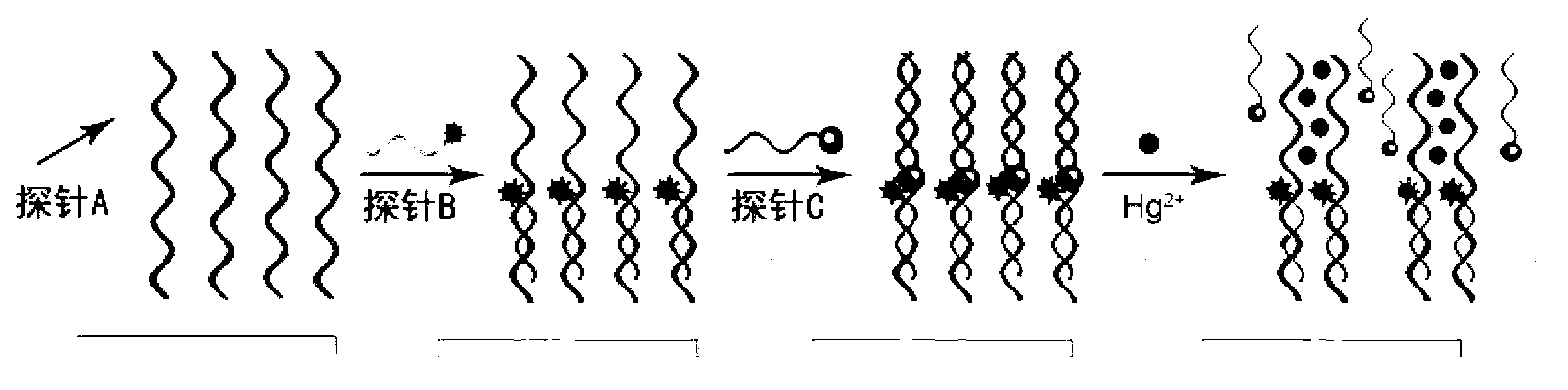
[0030] Embodiment 1: utilize probe A, probe B and probe C to prepare Hg 2+ Detection chip.

[0031] Probe A was made into a solution with a concentration of 10 μM, then mixed with the same volume of spotting solution, and arrayed on the surface of an aldehyde-modified glass slide with the microarray chip production system of Cartesian Company, placed at room temperature, 70% Store in relative humidity for 48-72h for fixation, then immerse the slide in 0.2% SDS at room temperature for a few minutes, then immerse in pure water for a few minutes, then immerse in 0.2% SDS twice, each time for 2min, then immerse in pure water Twice, 2min each time, dry. Hybridization solution (10mM MOPS, 100mM NaNO 3 ,pH7.2) Probe B was diluted to a final concentration of 2-5μM, dropped on the chip, covered with a cover slip, and hybridized overnight at room temperature. The next morning, place the chip at 4°C for 30 minutes, then at 25°C for 15 minutes. Then wash with 0.2% SDS, 2×SSC, 0.2×SSC ...

Embodiment 2

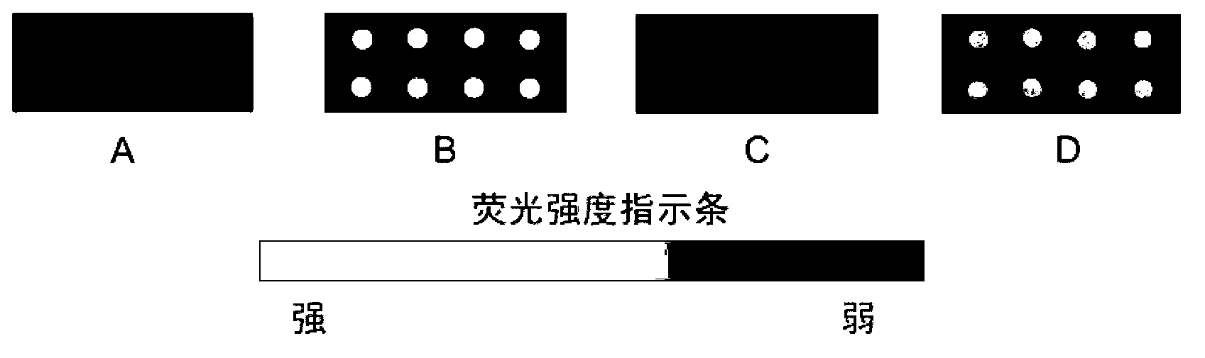
[0032] Example 2: Using probes A, B, and C to prepare a chip to detect 100nM Hg 2+ Solution, and step-by-step scanning of fluorescence intensity photos.

[0033] The chip preparation method is the same as that in Example 1, after the probe A is immobilized, after the probe B is hybridized, and after the probe C is hybridized, use the chip signal analysis system Scanarray 3000 of General Scanning Company to scan the photos ( figure 2 A-C). 10mM MOPS, 100mM NaNO 3 , pH7.2 diluted to prepare 100nM Hg 2+ solution, the Hg 2+ The solution is added to the prepared chip spots. After reacting at room temperature for 1 hour, take out the chip, and use 10mM MOPS, 100mM NaNO 3 , washed with pH7.2 buffer solution for 3 times, dried, and scanned with Scanarray 3000 chip signal analysis system of General Scanning Company ( figure 2 D).

[0034] The results showed that the chip had no fluorescence after probe A was immobilized; after hybridization of probe B, the fluorescence intensi...

Embodiment 3

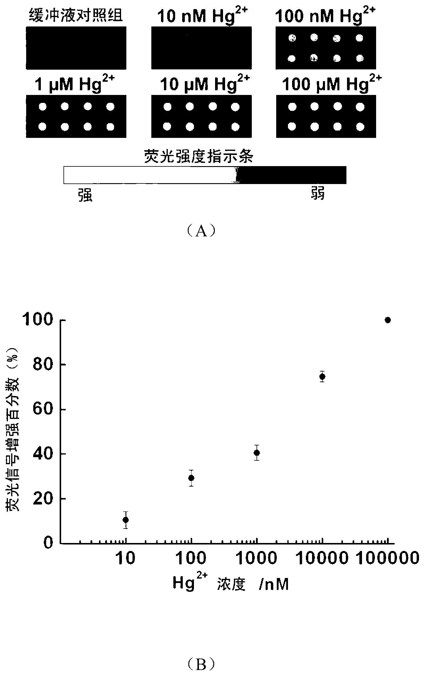
[0035] Embodiment 3: Utilize the chip prepared by probe A, B, C to detect different concentrations of Hg 2+ .

[0036] With 10mM MOPS, 100mM NaNO 3 , pH7.2 were diluted to prepare 10nM, 100nM, 1μM, 10μM, 100μM Hg 2+ , plus different concentrations of Hg 2+ The solution was placed on the prepared chip spots at room temperature and reacted for 1 h. With 10mM MOPS, 100mM NaNO 3 , washed 3 times with pH7.2 buffer and dried. The photos were scanned with the chip signal analysis system Scanarray 3000 of General Scanning Company ( image 3 A) and analyze the results ( image 3 B).

[0037] The results showed that in Hg 2+ In the presence of ions, the fluorescence intensity at the chip spot increases, and when Hg 2+ The higher the concentration, the greater the enhancement of fluorescence intensity. When Hg 2+ When the concentration is 100nM, compared with the fluorescence intensity of the buffer group, the fluorescence intensity at the spot is enhanced by 30%, with the inc...


PUM


Abstract

The invention relates to a fluorescence-enhanced Hg<2+> detection chip based on oligonucleotide chains and a method thereof. The invention is characterized in that Hg<2+> can specifically realize the covalent binding with T basic groups on two adjacent full thymine (T) oligonucleotide chains to form a stable intermolecular T-Hg<2+>-T structure, thus inducing the release of complementary chains hybridized with the full T oligonucleotide chains. The preparation method comprises the following steps: fixing a detection probe onto a chemically modified glass slide, and then respectively hybridizing with the fluorescence-labeled and quencher-labeled complementary chains. When the chip is in use, only a sample to be detected needs to be added onto the chip and kept for a time period, the chip is rinsed and then scanned by a fluorescence chip signal analysis system, and the change of a fluorescence signal is analyzed to realize Hg<2+> detection. The higher the Hg<2+> concentration in a sample is, the more the fluorescence signal is enhanced. The concentration range of the detected Hg<2+> is 10 nM-100 muM. The invention has favorable ionic selectivity.

Description

technical field [0001] The invention relates to a fluorescence-enhanced mercury ion detection chip and a method based on oligonucleotide chains, belonging to the technical field of biological analysis. Background technique [0002] Mercury is a highly toxic global environmental pollutant, especially its high mobility, persistence, methylation, bioaccumulation, and food chain amplification, even if it exists in a very small amount in the environment. The health of animals, plants and humans is also a great threat. The annual emission of mercury in the world is about 15,000 tons, mainly from mercury mines, metallurgy, chlor-alkali industry, electrical industry and combustion of fossil fuels. Mercury exists in various forms in the environment, water-soluble divalent mercury ions (Hg 2+ ) is one of the most common and stable forms of mercury contamination. [0003] How to effectively measure the content of mercury ions in the environment has become a problem facing the majori...

Claims


Application Information

Patent Timeline
IPC IPC(8): C12Q1/68G01N21/64
Inventor 赵建龙娄新徽杜娟徐元森
Owner SHANGHAI INST OF MICROSYSTEM & INFORMATION TECH CHINESE ACAD OF SCI
Why Eureka
Try Eureka