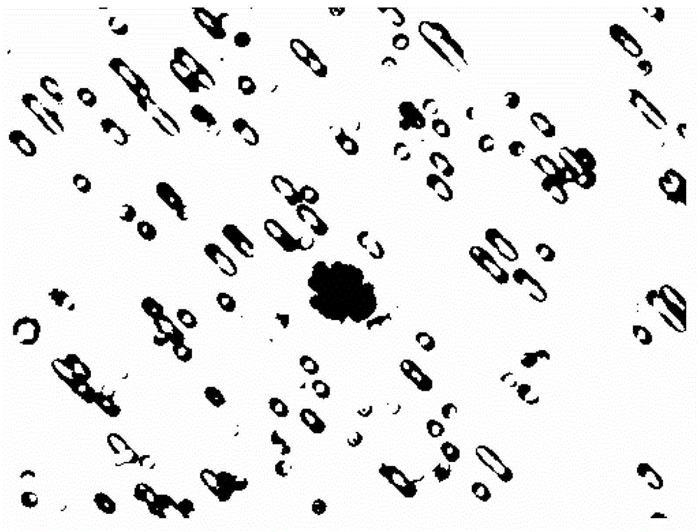
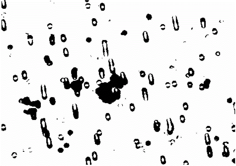
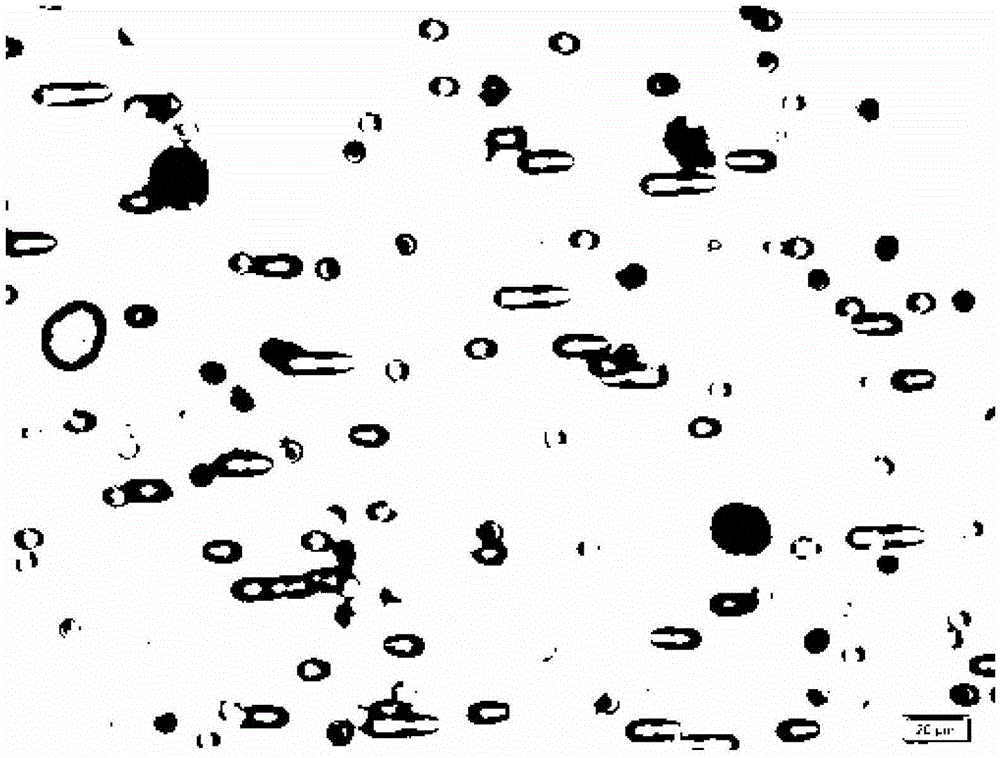
Circulating tumor cell dyeing kit and use thereof
A technology for dyeing reagents and tumor cells, which is applied in the fields of biotechnology and medicine, can solve problems such as complex procedures and long dyeing and identification operation cycles, and achieve the effects of simple preparation, true and reliable results, and improved work efficiency
- Summary
- Abstract
- Description
- Claims
- Application Information
AI Technical Summary
Problems solved by technology
Method used
Image
Examples
Embodiment 1
[0046] Preparation of circulating tumor cell staining kit:
[0047] 1. Preparation of basophilic staining solution:
[0048] Weigh 1000ml of analytical grade methanol with a graduated cylinder and add it to the beaker, weigh 1g of eosin Y with a balance, add it to the beaker, shake to fully dissolve the eosin Y, filter through a 0.22μm filter membrane, and store at room temperature for later use.
[0049] 2. Preparation of eosinophilic staining solution:
[0050] First weigh 10g of disodium hydrogen phosphate and 12.5g of potassium dihydrogen phosphate with a balance, dissolve them in 1000 ml of distilled water to make a PBS solution, then weigh 1g of medical melanin and 1g of azure I and dissolve them in the PBS solution. The pH was adjusted to 6.6, filtered through a 0.22 μm pore size filter, and stored at room temperature for later use.
[0051] 3. Sodium chloride solution preparation:
[0052] Weigh 1000ml of distilled water with a measuring cylinder and add it to the b...
Embodiment 2
[0058] Tumor cell line staining:
[0059] 1. Take 2ml of freshly passaged prostate cancer cell line PC3 and adjust the cell concentration to about 0.5×10 6 / ml, mix well.
[0060] 2. Use a pipette gun to take 1 μL (about 500 cells) of cells and add them to 25 ml of the fixative prepared in Example 1, and fix them for ten minutes.
[0061] 3. Wet the filter membrane of the filter with the sodium chloride solution prepared in Example 1.
[0062] 4. Transfer the above-mentioned fixative solution containing cells to the filter with a Pasteur tube, use negative pressure to make the fixative solution flow out through the filter membrane, and the cells are trapped on the filter membrane.
[0063] 5. Take a six-hole plate, place a piece of parafilm of appropriate size in one of the holes, and place the filter on the parafilm to prevent the liquid from flowing out of the filter.
[0064] 6. Add 300 μL of basophilic dye solution to the filter membrane, stain for 30 seconds, use negat...
Embodiment 3
[0070] Filter staining of blood samples from breast cancer patients:
[0071] 1. Use EDTA-2K anticoagulated blood collection tubes to collect 2.5ml of blood from breast cancer patients in clinical medical units.
[0072] 2. Centrifuge the blood, centrifugation conditions: 400G, 10 minutes; carefully suck off the supernatant with a pipette gun.
[0073] 3. Transfer the above blood to 25ml fixative with a pipette gun, and fix it for 10 minutes.
[0074] 4. Wet the membrane in the filter with 5% (w / v) glucose solution.
[0075] 5. Transfer the blood-containing fixative to the filter with a Pasteur tube, and use negative pressure to make it flow out through the filter. Larger cells in the blood and circulating tumor cells are trapped on the membrane.
[0076] 6. Add an appropriate amount of 5% glucose solution to rinse, use negative pressure to make it flow out, and try to remove it as clean as possible.
[0077] 7. Take a six-hole plate, put a piece of sealing film of appropri...
PUM
| Property | Measurement | Unit |
|---|---|---|
| concentration | aaaaa | aaaaa |
| pore size | aaaaa | aaaaa |
Abstract
Description
Claims
Application Information
 - R&D
- Intellectual Property
- Life Sciences
- Materials
- Tech Scout
- Unparalleled Data Quality
- Higher Quality Content
- 60% Fewer Hallucinations
Browse by: Latest US Patents, China's latest patents, Technical Efficacy Thesaurus, Application Domain, Technology Topic, Popular Technical Reports.
© 2025 PatSnap. All rights reserved.Legal|Privacy policy|Modern Slavery Act Transparency Statement|Sitemap|About US| Contact US: help@patsnap.com


