
Multifunctional tissue cell stereology quantitative analysis slide system

A tissue cell quantitative analysis technology, applied in the field of medical and health products, can solve the problems that the geometric parameters of tissue cells cannot be directly judged under the microscope, the process of visual field capture cannot be accurately repeated, and the difficulty of positioning and judgment increases, so as to reduce proficiency requirements, achieve repeatability, and reduce the effect of human judgment

Active Publication Date: 2013-04-03
THE THIRD AFFILIATED HOSPITAL OF THIRD MILITARY MEDICAL UNIV OF PLA
View PDF5 Cites 4 Cited by
  • Summary
  • Abstract
  • Description
  • Claims
  • Application Information

AI Technical Summary

Problems solved by technology

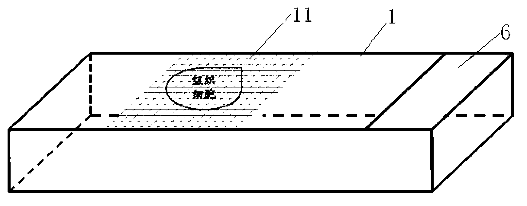
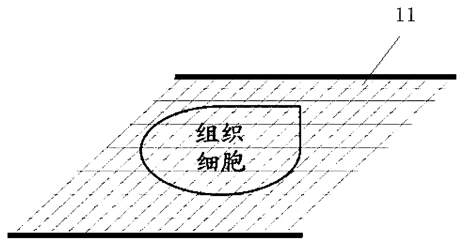
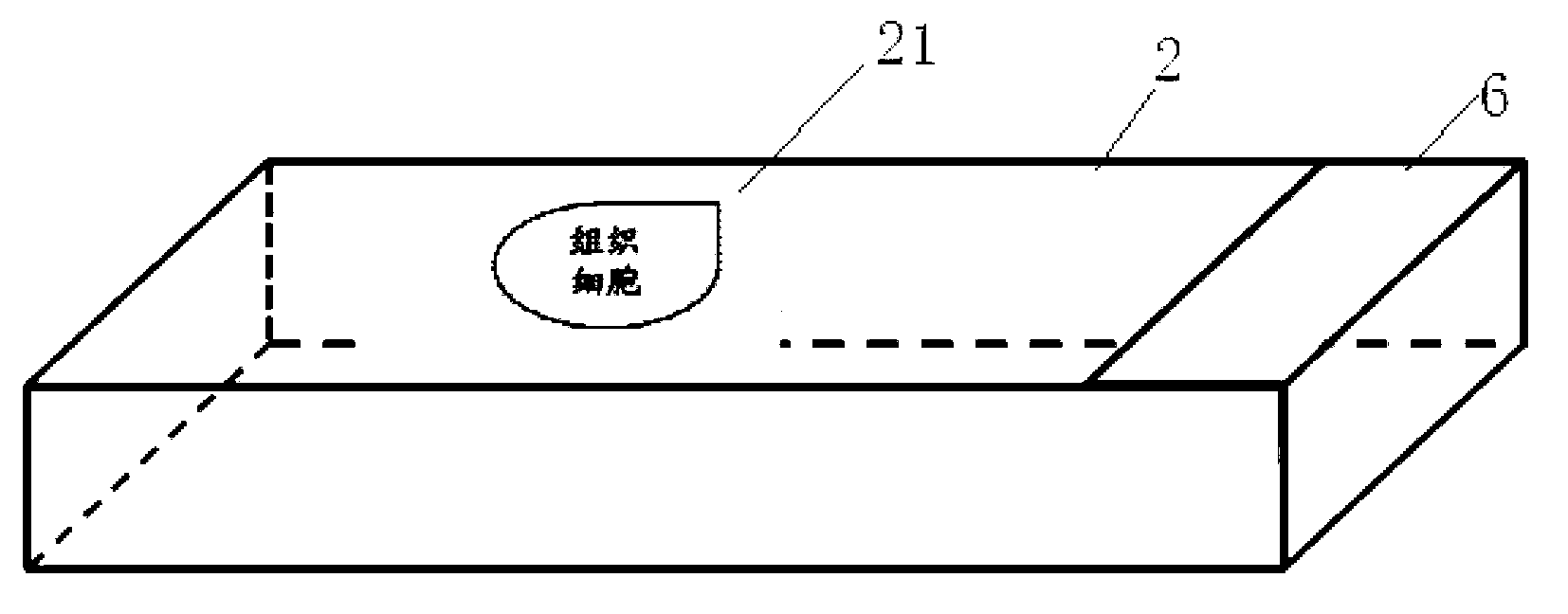
[0004] In the process of quantitative analysis of tissue cells, it is impossible to accurately locate under the microscope, the process of capturing the field of view cannot be accurately repeated, the geometric parameters of tissue cells cannot be directly judged under the microscope, and the difficulty of the above-mentioned positioning and judgment is further increased under dark field conditions. The present invention A set of multifunctional tissue cell stereological quantitative analysis slide system is proposed, which can realize the precise positioning of two-dimensional planes in the process of qualitative, semi-quantitative and quantitative evaluation of tissue cells under normal field and dark field conditions, as well as tissue cell Geometric parameters are directly judged under the microscope

Method used


Image

  • Multifunctional tissue cell stereology quantitative analysis slide system
  • Multifunctional tissue cell stereology quantitative analysis slide system
  • Multifunctional tissue cell stereology quantitative analysis slide system

Examples


example 1

[0036] Example 1: Quantitative analysis of cultured cell morphology

[0037] 1. Under the ordinary or fluorescence microscope / confocal laser scanning microscope, accurately locate a single target cell, and calculate the number of target cells according to the stereological method.

example 2

[0038] Example 2: Analysis of Migration Function of Cultured Cells

[0039] 1. Observing the directional migration speed of cells or target cells in tissues under the action of bioelectric field or chemotaxis under ordinary or fluorescence microscope / confocal laser scanning microscope, and studying the directional movement function of cells by evaluating the migration activity of cells.

example 3

[0040] Example 3: Quantitative analysis of tissue cell morphology

[0041] 1. Under the ordinary or fluorescence microscope / confocal scanning microscope, accurately locate the tissue cells, and calculate the number of target cells in a specific area of ​​the tissue according to the stereological method.


PUM


Abstract

The invention relates to a multifunctional tissue cell stereology quantitative analysis slide system. The slide system comprises a tissue section area analysis slide, a tissue cavity structure analysis slide, a target spot tissue cell structure analysis slide, a tissue cell quantitative analysis slide, and a body fluid cell quantization slide. On the basis of conventional magneification factor (40-400) of an inverted phase contrast optical microscope, stereology quantitative analysis function areas of corresponding slides are divided by linear grooves of different intervals (5mum and 10mum) or grooves with arc-shaped radius intervals of 5mum, and each groove is 1-2mum deep; on the basis of meeting the observation of a light source of the common optical microscope, when slides are observed in dark field, colourless phthalic ester composite fluorescent materials of different fluorescence color developing effects are arranged in depression areas, thereby forming a set of complete tissue cell stereology quantitative analysis slide system. The multifunctional tissue cell stereology quantitative analysis slide system can realize precise positioning of a two-dimensional plane in tissue cell qualitative process, semiquantitative process and quantitative evaluation process under the conditions of common field and dark field, as well as direct judgment on geometric parameters of tissue cells under the microscope.

Description

technical field [0001] The invention relates to the technical field of medical and sanitary products, in particular to a quantitative analysis tool using histology and cell stereology. Background technique [0002] With the rapid development of modern pathology technology, human's understanding of disease etiology, pathology, diagnosis and treatment has reached the level of tissue cells. The microscope invented by the Dutch at the end of the 16th century was a milestone in promoting human understanding of the microscopic world of diseases, while the electron microscope invented by E. Ruska and M. Knoll in 1932 further deepened the observation of tissue cells to the subcellular level. In this process, human observation of tissue cells or subcellular structures relies on various classical staining (such as hematoxylin-eosin, crystal violet, malachite green) and immunostaining (such as antibodies against specific antigens of tissue cells, intracellular RNA Specific binding fra...

Claims


Application Information

Patent Timeline
IPC IPC(8): C12M1/34G02B21/34
Inventor 杨策蒋建新李海胜王海燕杜娟高洁王正国
Owner THE THIRD AFFILIATED HOSPITAL OF THIRD MILITARY MEDICAL UNIV OF PLA
Learn More