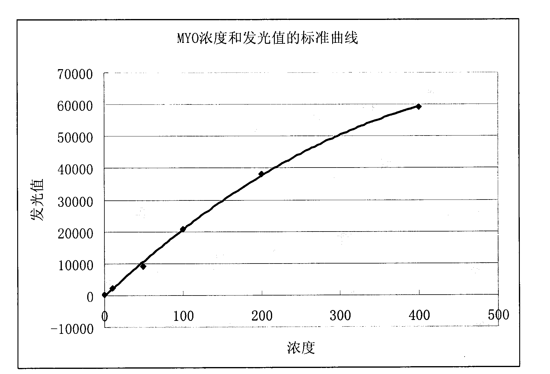
Myoglobin enzymatic chemiluminescence immunodetection method and kit
A human myoglobin and detection method technology, applied in the human myoglobin enzymatic chemiluminescence immunoassay method and kit, the field of enzymatic chemiluminescence detection and analysis of human myoglobin, can solve the problem that there is no chemiluminescence analysis method, measurement Human myoglobin research and application, to achieve the effect of small batch-to-batch difference, improved sensitivity and specificity, and high specificity
- Summary
- Abstract
- Description
- Claims
- Application Information
AI Technical Summary
Problems solved by technology
Method used
Image
Examples
Embodiment 1
[0043] Embodiment 1 Preparation of horseradish peroxidase (HRP) labeled antibody
[0044] Concentrate the anti-human myoglobin monoclonal antibody to 2mg / mL. Use the modified sodium iodate method for labeling, the specific steps are:
[0045] (1) 5mg of HRP (purchased from Sigma, USA) was dissolved in 0.5ml double distilled water, and newly prepared 0.06Mol / L NaIO was added 4 0.5ml aqueous solution, mix well, set at 4°C for 30min;
[0046] (2) Add 0.5ml of 0.16Mol / L ethylene glycol aqueous solution, and let stand at room temperature for 30min;
[0047] (3) Add 2.5ml of an aqueous solution containing 5mg of purified anti-human myoglobin monoclonal antibody (clone number 7001), mix well, put it into a dialysis bag, and slowly stir and dialyze against 0.05Mol / L pH 9.5 carbonate buffer 6h (or overnight), make it combine;
[0048] (4) Take out the solution in the dialysis bag, add NaBH 4 Solution (5mg / ml) 0.2ml, mix well, set at 4°C, 2h;
[0049] (5) Slowly add an equal volum...
Embodiment 2
[0051] Example 2 Adsorption immobilization of coated antibody
[0052] Dilute anti-human myoglobin monoclonal antibody (clone number 7004) to 2.5 μg / ml with 0.02M, pH7.4 phosphate buffer solution (PBS solution), add polystyrene microwell plate, 100 μL / well, 4 °C Overnight, wash three times with washing solution, block with 300 μL / well of 1% bovine serum albumin at room temperature for 2 hours, and then pat dry for later use.
[0053] The preparation method of washing liquid is: sodium chloride (NaCl) 2g, sodium dihydrogen phosphate (NaH 2 PO 4 .12H 2 O) 2.9g, dipotassium hydrogen phosphate (K 2 HPO 4 ) 0.2g, Potassium Chloride (KCl) 0.2g, Triton X-100 0.5mL, Tween-20 0.5mL, Bronidox-L 5g, make 1000mL with purified water, use Filter through a 0.2 μm filter and store at room temperature.
Embodiment 3
[0054] Example 3 Affinity immobilization of coated antibodies
[0055] Dilute avidin with 0.02M, pH7.4 phosphate buffer solution to 1.0 μg / ml, add to a polystyrene microwell plate, 100 μL / well, overnight at 4°C, wash with washing solution (preparation method is the same as in Example 2) Three times, add biotin-labeled anti-human myoglobin antibody (clone number 7004) (see Thermofisher for biotin labeling method) Instructions for use of Sulfo-NHS-Biotin, product number is 21217), concentration 1.0 μg / ml, 100 μL / well, overnight at 4°C, washed three times with washing solution, blocked with 1% bovine serum albumin 300 μL / well at room temperature for 2 hours, then photographed Dry and set aside.
PUM
 Abstract
Description
Claims
Application Information
 - R&D
- Intellectual Property
- Life Sciences
- Materials
- Tech Scout
- Unparalleled Data Quality
- Higher Quality Content
- 60% Fewer Hallucinations
Browse by: Latest US Patents, China's latest patents, Technical Efficacy Thesaurus, Application Domain, Technology Topic, Popular Technical Reports.
© 2025 PatSnap. All rights reserved.Legal|Privacy policy|Modern Slavery Act Transparency Statement|Sitemap|About US| Contact US: help@patsnap.com


