Micro-fluidic chip capable of producing stable concentration gradient and cell co-culture method

A microfluidic chip and concentration gradient technology, applied in the field of biomedical research, can solve the problems of large amount of cells, large consumption of reagents, lack of biological information, etc., and achieve important biomedical research value and economic value, sample amount Few, easy-to-use effects

Inactive Publication Date: 2013-05-08
DALIAN INST OF CHEM PHYSICS CHINESE ACAD OF SCI
View PDF4 Cites 25 Cited by
  • Summary
  • Abstract
  • Description
  • Claims
  • Application Information

AI Technical Summary

Problems solved by technology

The above three methods all have many disadvantages, such as large consumption of reagents, large amount of cells, inability to conveniently realize fluid control, etc.
At the same time, it is difficult to simula

Method used


Image

  • Micro-fluidic chip capable of producing stable concentration gradient and cell co-culture method
  • Micro-fluidic chip capable of producing stable concentration gradient and cell co-culture method
  • Micro-fluidic chip capable of producing stable concentration gradient and cell co-culture method

Examples


Embodiment 1

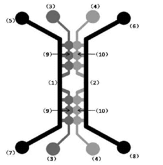
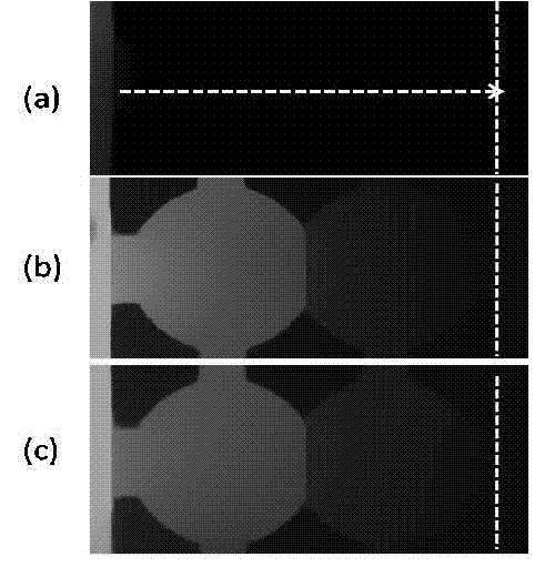
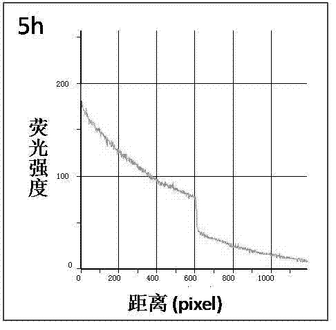
[0021] The microfluidic chip used was designed and manufactured by our laboratory. The chip is formed by irreversible sealing of upper and lower layers, the upper layer material is PDMS polymer, and the lower layer material is glass. Such as figure 1 As shown, the size of the perfusion channel is 10 mm in length, 500 μm in width, and 200-300 μm in height (the black area in the figure), the size of the culture chamber A is 1 mm in diameter, and 120 μm in height (the gray area in the figure), and the culture chamber B The dimensions are 1 mm in diameter and 50 μm in height (light gray area in the figure). Take 5 μl of three-dimensional Matrigel with a pipette, add it to culture chambers B and A sequentially through the sample inlet pool, and place them in a 37°C incubator for 20-30 min. After gelling, the culture solution containing FITC-dextran and the culture solution without fluorescent dye were added to the perfusion channels A and B respectively, and the continuous perfus...

Embodiment 2

[0024] Cell contact co-culture: including four parts: live cell labeling, cell loading, cell chip co-culture and photo detection. First, CellTracker Red and CellTracker Green were used to stain salivary adenoid cystic carcinoma cells ACCM and mesenchymal stem cells MSCs, respectively, with a final concentration of 5 μM. After the staining was completed, the cells were digested, and the mixture of salivary adenoid cystic carcinoma cells ACCM and matrigel was added to the culture chamber B through the sample inlet pool B, and placed in CO 2 In the incubator; after gelling for 20-30 min, add mesenchymal stem cells MSCs into the culture chamber A through the sample inlet pool A; place in CO 2 In the incubator; after gelling for 20-30 min, add cell culture medium into perfusion channels A and B, and replace the cell culture medium once a day. Photographs were taken at 0 h, 24 h, and 48 h to record the location of the cells, and the initial intersection of the two cells was taken a...

Embodiment 3

[0026] Directed migration of tumor cells: including live cell labeling, cell loading, cell chip culture, generation of chemokine concentration gradients, and cell photography. First, CellTracker Red was used to stain salivary adenoid cystic carcinoma cells ACCM in vivo, and the final concentration was 5 μM. After staining, proceed to cell digestion. Salivary gland adenoid cystic carcinoma cells ACCM were mixed in matrigel three-dimensional matrigel, and the mixture of cells and matrigel was added through the sample inlet pool B, and placed in CO 2 In the incubator, after 20-30 minutes of gelation, add matrigel without any cells through the sample inlet pool A. After 20-30 minutes of gelation, a clear interface between the gel and cells can be formed, and then the perfusion is performed through the syringe pump. Channels A and B respectively introduce the culture solution containing chemokines and the culture solution not containing chemokines, and ensure continuous perfusion ...


PUM


Abstract

The method provides a micro-fluidic chip capable of producing stable concentration gradient and a cell co-culture method. The micro-fluidic chip is composed of a perfusion channel, an inlet pool, a culture chamber, a waste liquid pool and a micro channel. Direct coculture and mixed coculture of cells can be carried out on a chip with an area of few square centimeters, and at the same time, the stable concentration gradient of cell chemotactic factor can be maintained, so that a micro environment of tumor cells is constructed, and mutual effects between biological cells and cytokines, between cells and matrixes, between cells can be simulated effectively. The micro-fluidic chip has the advantages of simple operation, simple manufacture, small sample usage and the like, and has significant biomedical research values and economic values.

Description

technical field [0001] The invention belongs to the field of applying microfluidic chip technology to biomedical research, and in particular relates to a microfluidic chip and a cell co-cultivation method for generating stable concentration gradients. Background technique [0002] Cell biology research is developing from the observation and study of a single cell to the observation and study of the interaction of two or even multiple types of cells. For complex biological systems, a system in which multiple cells exist at the same time can better simulate the real cellular microenvironment, thereby more comprehensively and deeply exploring the complex network between cells and the signaling pathways that mediate their interactions, etc. , for the study of body development, inflammatory response, immune response, organ function and tumorigenesis mechanism are very important. Cell co-culture is a common method for studying cell-cell interactions. Traditionally, cell co-cult...

Claims


Application Information

Patent Timeline
IPC IPC(8): C12M3/00C12N5/09C12N5/0775
Inventor 秦建华马慧朋
Owner DALIAN INST OF CHEM PHYSICS CHINESE ACAD OF SCI
Why Eureka
Try Eureka