
Human tumor invasion and metastasis histioid in vitro three-dimensional model and construction and evaluation thereof

A three-dimensional model, metastatic technology, applied in the direction of tumor/cancer cell, microorganism determination/inspection, biochemical equipment and methods, etc. problem, to achieve the effect of good biocompatibility, maintaining cell viability, and reducing sensitivity

Inactive Publication Date: 2013-06-19
DALIAN INST OF CHEM PHYSICS CHINESE ACAD OF SCI
View PDF1 Cites 14 Cited by
  • Summary
  • Abstract
  • Description
  • Claims
  • Application Information

AI Technical Summary

Problems solved by technology

In the in vitro tumor invasion and metastasis model, the traditional plane-cultured metastatic human tumor cell lines have been unable to meet the experimental analysis of the molecular pathological mechanism of tumor invasion and metastasis, and the expression and behavior of a large number of plane-cultured cell lines reported so far are similar to those of in vivo cancer cells. The actual pathological conditions of the cells are quite different [references: Pampaloni, F., E.G.Reynaud, et al. (2007). "The third dimension bridges the gap between cell culture and live tissue." Nature Reviews Molecular Cell Biology8 (10): 839-845.], while the three-dimensional cultured histiocyte-like spheres are closer to the expression and behavior of cells in the tumor microenvironment in vivo, and because the in vitro three-dimensional culture is easy to operate on a large scale, the in vitro three-dimensional model is used to explore and establish new in vitro invasion Metastasis models or metastasis research systems can provide guidance for new theories and mechanisms of tumor metastasis
[0003] The traits of traditional planar cultured cells are uniform and non-polar, and the cell shape affects the cytoskeleton, thereby affecting the secretory function of cells and the abnormal expression of some key genes, and planar cultured cells cannot reflect the interaction between cells and between cells and the matrix. In vitro simulation studies on biological processes such as intercellular adhesion, signal transduction, cell movement, invasion and metastasis, etc.

Method used


Image

  • Human tumor invasion and metastasis histioid in vitro three-dimensional model and construction and evaluation thereof
  • Human tumor invasion and metastasis histioid in vitro three-dimensional model and construction and evaluation thereof
  • Human tumor invasion and metastasis histioid in vitro three-dimensional model and construction and evaluation thereof

Examples


Embodiment 1

[0032] An in vitro three-dimensional model of human tumor invasion and metastasis tissue-like, the specific steps are:

[0033] Step 1: Put 2×10 6 Human liver cancer cells were suspended in 1ml sodium alginate solution (15g / L), and the droplets were sprayed into 1.0mol / L CaCl by electrostatic droplet method 2 solution, and reacted for 20 minutes to obtain calcium alginate gel microspheres with a particle size of 450 μm;

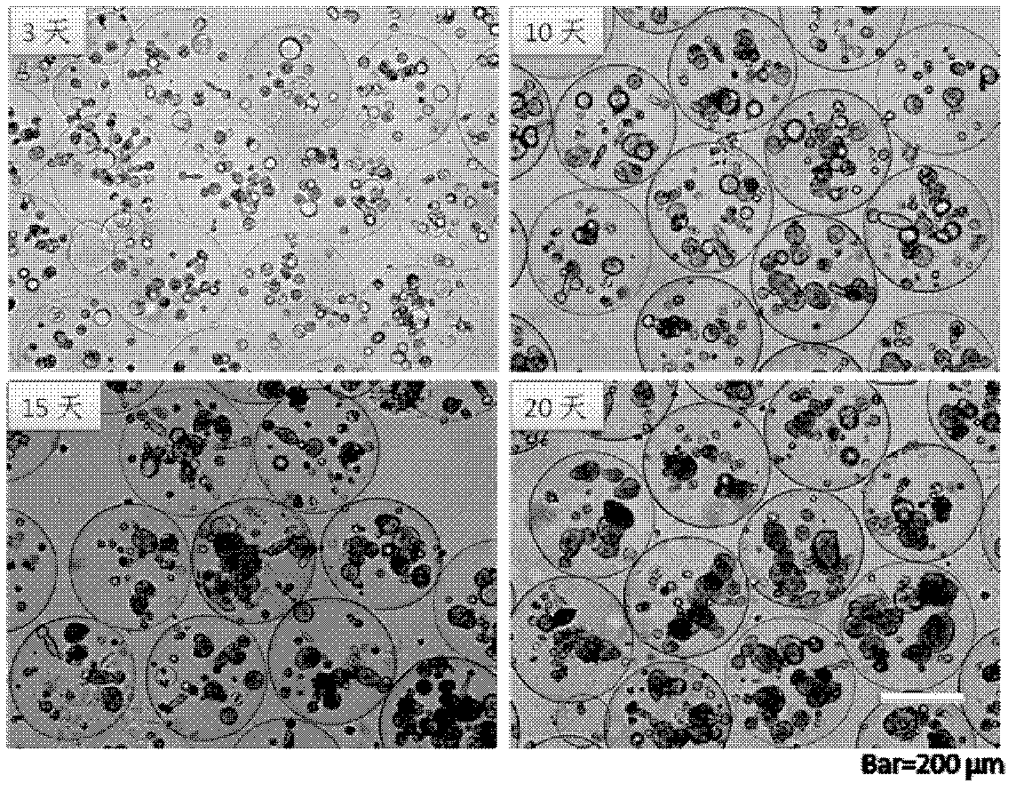
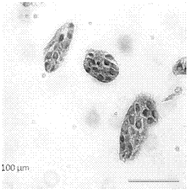
[0034] The second step: the above-mentioned hydrogel microspheres were naturally settled for 4 minutes, and then inoculated into high-glucose DMEM medium (GIBCO company) containing 10% fetal bovine serum (GIBCO company) according to the volume of 10% after the sedimentation, and after standing for 5 minutes, At 37°C, with a volume fraction of 5% CO 2 Cultured in an incubator for 20 days under air-saturated humidity conditions, tumor-like organoids with a diameter of 150 μm can be obtained, such as figure 1 Shown is a map of cell-like tissue formed by liver...

Embodiment 2

[0042] An in vitro three-dimensional model of human tumor invasion and metastasis tissue-like, the specific steps are:

[0043] Step 1: Put 5×10 5 Human breast cancer cells were suspended in 1ml sodium alginate solution (10g / L), and the droplets were sprayed into 1.0mol / L CaCl by electrostatic droplet method 2 solution, and reacted for 30 minutes to obtain calcium alginate gel microspheres with a particle size of 300 μm;

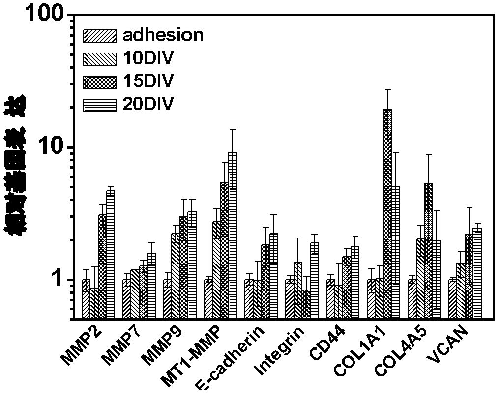
[0044] The second step: the above-mentioned hydrogel microspheres were naturally settled for 6 minutes, and then inoculated into the RPMI1640 medium (GIBCO company) containing 10% fetal bovine serum (GIBCO company) according to the volume of 10% after the sedimentation, after standing for 5 minutes, at 37 °C, containing 5% CO by volume 2 The tumor-like organoids with a diameter of 100 μm can be obtained by culturing in an incubator for 15 days under air-saturated humidity conditions.

[0045] Experiment to verify the protein expression of tumor invasion a...

Embodiment 3

[0047] An in vitro three-dimensional model of human tumor invasion and metastasis tissue-like, the specific steps are:

[0048] Step 1: Put the 8×10 5 Human head and neck squamous cell carcinoma cells were suspended in 1ml sodium alginate solution (40g / L), and the droplets were sprayed into 0.1mol / L CaCl by electrostatic droplet method 2 solution, and reacted for 40 minutes to obtain calcium alginate gel microspheres with a particle size of 800 μm;

[0049] The second step: the above-mentioned hydrogel microspheres were naturally settled for 3 minutes, and were inoculated into the RPMI1640 medium (GIBCO company) containing 8% fetal bovine serum (GIBCO company) according to the volume of 10% after the sedimentation, after standing for 3 minutes, at 37 °C, containing 5% CO by volume 2 The tumor-like organoids with a diameter of 50 μm can be obtained by culturing in an incubator for 15 days under air-saturated humidity conditions.

[0050] In vitro tumor invasion and metastasi...


PUM

PropertyMeasurementUnit
Diameteraaaaaaaaaa
Diameteraaaaaaaaaa
Particle sizeaaaaaaaaaa

Abstract

The invention discloses an in vitro model simulating human tumor metastasis. Cells in the model have a tumor high metastasis character. The model is constructed by building of a micro spherical hydrogel support frame by tumor high metastasis cells, building of a tumor cell in vitro metastasis model and identifying of the constructed tumor cell in vitro metastasis model. The model can grow in a three-dimensional culturing cell in a clustering mode, and tumor cell biological behaviors in metastasis can be detected. The model has important value in studying of a clinical tumor metastasis mechanism and controlling of screening of metastasis medicine and can be taken as a technical platform for manufacturing anti-tumor metastasis gene vaccines and building screening of anti-tumor metastasis medicine.

Description

technical field [0001] The invention provides an in vitro three-dimensional tissue-like model of human tumor invasion and metastasis, and belongs to the technical field of tumor invasion and metastasis in vitro models. Background technique [0002] The process of tumor metastasis is complex and difficult to control, and 90% of the mortality of clinical cancer patients is caused by tumor metastasis. To some extent, preventing tumor metastasis can control the death caused by tumor. Tumor invasion and metastasis are malignant biological behaviors of tumor cells. Tumor invasion is also known as direct spread of tumors, and the process of discontinuous dissemination of tumor cells and their growth in distant sites is metastasis. The above-mentioned process is a complex and multi-step process, which generally includes detachment of tumor cells from the primary tumor focus; degradation of the basement membrane, outward infiltration, migration and adhesion to vascular endothelial c...

Claims


Application Information

Patent Timeline
IPC IPC(8): C12N5/09C12N11/10C12N11/04C12Q1/02
Inventor 马小军徐小溪孙广炜于炜婷王为李楠刘畅
Owner DALIAN INST OF CHEM PHYSICS CHINESE ACAD OF SCI
Learn More