Fluorescent staining kit for rapid detection on biological cell viability, and application of same

A biological cell, fluorescent dyeing technology, applied in measurement devices, particle and sedimentation analysis, individual particle analysis, etc., to achieve the effects of low toxicity, long exclusion culture period, and ease of use

Active Publication Date: 2013-06-26
DONGHUA UNIV
View PDF4 Cites 13 Cited by
  • Summary
  • Abstract
  • Description
  • Claims
  • Application Information

AI Technical Summary

Problems solved by technology

The disadvantage is that because the sample to be tested is often not easily dispersed into a single cell, the single colony formed after culture may come from 2-3 or more cells in the sample, or due to the different growth cycles of the bacteria, in the same species Due to the difference in adaptability in the medium, some bacteria need 3 days to grow colonies, and some bacteria need 1 week or even longer, so the results of plate colony counts are often low, and colony formation is usually used. Units (colony-formingunits, cfu) instead of the absolute number of colonies to express the live bacteria content of the sample

Method used


Image

  • Fluorescent staining kit for rapid detection on biological cell viability, and application of same
  • Fluorescent staining kit for rapid detection on biological cell viability, and application of same
  • Fluorescent staining kit for rapid detection on biological cell viability, and application of same

Examples


Embodiment 1

[0038] (1) Anthocyanin dyes are dissolved in dimethyl sulfoxide (DMSO) to a concentration of 5-6mM, which is the prepared concentrated solution of anthocyanin dyes for storage.

[0039](2) PI dye was dissolved in ultrapure water to 100 μg / mL, and stored away from light

[0040] (3) Pipette 1mL of Acetobacter xylinum solution into a 2mL centrifuge tube wrapped with tinfoil.

[0041] (4) Add 300 μL of anthocyanin dye diluted 1000 times with buffer and 200 μL of 100 μg / mL PI dye, shake up and down several times, and place in the dark for 10-30 minutes. The dilution buffer is 1×TE buffer (containing 10 mM Tris-HCl buffer in 1 mM EDTA, pH 8.0).

[0042] (5) Filter the stained bacterial solution through a 0.2 μm black filter membrane (Whatman), trap the bacteria on the black filter membrane, transfer the filter membrane to a glass slide, drop a drop of non-fluorescent essential oil in the center of the membrane, Cover with a coverslip.

[0043] (6) Observe the slide under the 100...

Embodiment 2

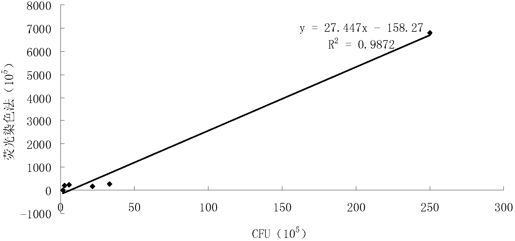
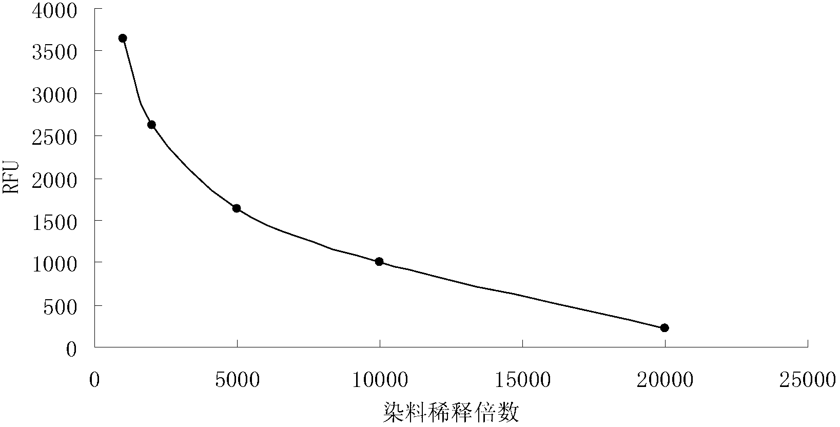
[0045] (1) Dilute the concentrated solution of anthocyanin dye storage by 1000 times to 20000 times, use dyes with the same volume but different dilution times to stain the bacteria according to steps 1-3 in Example 1, and use Molecular Devices Flexstation II calcium flow Fluorescence instrument was used to test, and it was found that the fluorescence intensity of the bacteria decreased with the increase of the dilution factor of the dye. When the dilution was 5000 times, the dyeing effect was better and the amount of dye was relatively low, which was more economical. If the dilution factor increases, the detected fluorescence value is close to the range of the error limit, and the error of the detection value increases (experimental results such as figure 2 ).

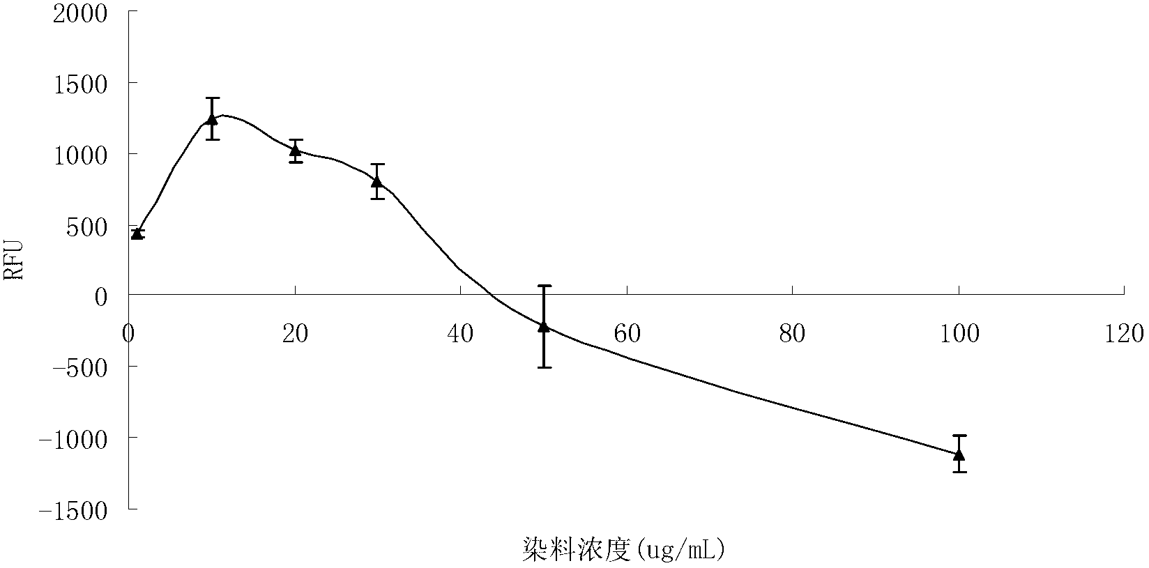
[0046] (2) The concentration of the dye PI was prepared from 1 μg / mL to 100 μg / mL, and the dyes with the same volume but different dilution times were used to stain the bacteria. It was found that the dyeing effect w...

Embodiment 3

[0048] (1) Cultivate Staphylococcus aureus until the turbidity OD value of the bacterial concentration is 0.7, take 25mL of the bacterial liquid and centrifuge at 12000r / min for 10-15 minutes;

[0049] (2) The collected bacteria were resuspended in 2 mL of normal saline;

[0050] (3) Dissolve 1mL of bacterial solution in 20mL of normal saline, and another 1mL in 20mL of 70% isopropanol;

[0051] (4) Stir every 15 minutes and incubate at room temperature for 1 hour;

[0052] (5) Centrifuge at 12000r / min for 10-15 minutes;

[0053] (6) Resuspend in 20mL of normal saline, then centrifuge and wash, the operation steps are the same as above;

[0054] (7) Resuspend with 10mL of normal saline respectively, and mix live bacteria and dead bacteria according to the ratio in Table 1;

[0055] Table I

[0056] Ratio of live bacteria to dead bacteria

[0057] (8) Add 140 μL of the mixed bacterial solution to the microwell plate, add 40 μL of 5 mM anthocyanin dye and 20 μL of ...


PUM


Abstract

The invention relates to a fluorescent staining kit for rapid detection on biological cell viability, and application of the same. The kit comprises an anthocyanin dye and a propidium iodide dye. The detection comprises the following steps of: adding the anthocyanin dye and the propidium iodide in the kit are added in cell suspension, observing and counting the cells by a fluorescence microscope or detecting the cells by a microporous fluorescent plate reader under wavelength of emitted light, and reflecting the viability of the cells in biologic sample liquid according to the strength of fluorescence values of different colors. The kit can be used for rapidly observing or detecting the amount of living cells and the amount of dead cells of various cells in the sample in real time, is visual and handy, eliminates the inference of other substances and the defect of long culture cycle, and is not only suitable for animal cells, but also suitable for most gram negative bacteria and gram positive bacteria.

Description

technical field [0001] The invention belongs to the field of cell counting, in particular to a fluorescent staining kit for rapidly detecting the life and death state of biological cells and its application. Background technique [0002] At present, the methods for determining the number of living cells include optical microscope reading counting method, plate coating culture colony counting method, photoelectric turbidimetric method, maximum probability method, and membrane filtration method. [0003] Microscopic direct counting method: Place a small amount of the suspension of the sample to be tested on a special glass slide (also known as a bacteria counter) with a definite area and volume, and directly observe and count under a microscope. The method is simple, fast and intuitive, and can be used for counting suspensions such as yeast, bacteria, mold spores and animal cells. The disadvantage is that it cannot accurately identify the dead and alive state of cells, and th...

Claims


Application Information

Patent Timeline
Patent Type & Authority Applications(China)
IPC IPC(8): G01N15/10
Inventor 洪枫张硕杜倩雯兰水陈琳李登新
Owner DONGHUA UNIV
Why Eureka
Try Eureka