Method for promoting epidermal cell proliferation
A kind of epidermal cells, low expression technology, applied in the field of stem cells and genetic engineering
- Summary
- Abstract
- Description
- Claims
- Application Information
AI Technical Summary
Problems solved by technology
Method used
Image

Examples
Embodiment 1
[0077] Example 1: Construction of the lentiviral overexpression plasmid pGC-FU-JAM1-GFP to infect human epidermal cells (hMSC).
[0078] 1. Separation and culture of epidermal cells
[0079] Adult body skin comes from Plastic Surgery of Changhai Hospital. Epidermal cells are obtained by primary culture.
[0080] 2. Preparation of total RNA of human epidermal cells
[0081] Using the total RNA extraction kit from Shanghai Huashun Biological Engineering Co., Ltd., the total RNA of human epidermal cells was extracted according to the conventional guanidine isothiocyanate method. Methods as below:
[0082] Take a monolayer of epidermal cells grown in a culture dish with a diameter of 3.5 cm, discard the medium directly, and add 1 ml of TRIzol to dissolve the cells. After the cells are fully dissolved, remove the cell lysate with a pipette. Incubate the cell lysate sample at 15~30°C for 5 minutes to completely decompose the ribosome. Add 0.2ml of chloroform per 1ml of TRIzol, close the c...
Embodiment 2
[0143] Example 2: Cell experiment (in vitro experiment)
[0144] Use fluorescence microscope to take pictures, draw growth curves, immunocytochemistry, western-blot and other biological experiment methods to analyze cell morphology changes, target gene expression and cell surface marker keratin expression from various aspects such as cell morphology and protein expression.
[0145] The specific method is as follows:
[0146] 1) Comparison of cell growth status
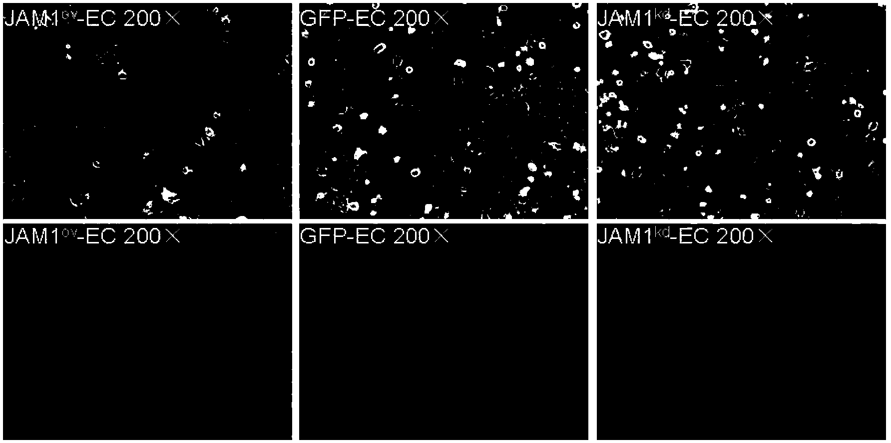
[0147] Observation of JAM1 infected by lentivirus with an inverted fluorescence microscope ov -EC, JAM1 kd -EC and GFP-EC. No obvious changes in cell morphology, such as figure 1 Shown.
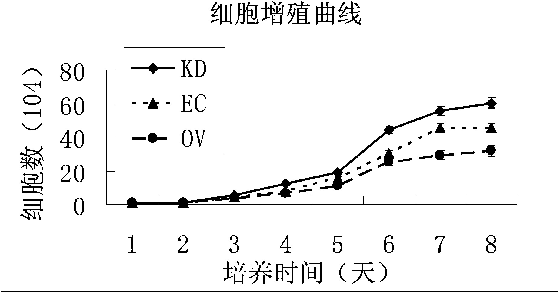
[0148] 2) Draw a growth curve to detect cell proliferation
[0149] JAM1 ov -EC, JAM1 kd -EC and GFP-EC were seeded on a 24-well plate with 10,000 cells per well, cultured for 7 days, trypsinized into a single cell suspension every day, and carried out cell counting experiments with the help of a cell counting plate and a microscope, each ty...
Embodiment 3
[0191] Example 3: In vivo tumorigenicity test
[0192] The nude mouse BALB / c Nu strain used in the experiment, SPF grade. Weighing about 15~25g, 3 weeks old, purchased from Shanghai Experimental Animal Center. A total of 12 nude mice, divided into 4 groups: JAM1 ov -EC injection group, GFP-EC injection group, JAM1 kd -EC injection group, PBS injection group. Cell volume is 10 4 Use a 1ml syringe to draw 0.15ml of cell suspension and inject it under the skin of the back of the forelimb of nude mice. They were fed under SPF-level conditions, observed daily, and harvested 4 weeks after transplantation.
[0193] Animals in each group showed no difference in appearance and activity after experimental treatment. There was no difference in H-E staining, liver, spleen, kidney and other organs among nude mice in each group after sacrifice.
[0194] H-E staining was performed on the skin tissues of the injection sites of nude mice in each group. Observation under the microscope revealed t...
PUM
 Abstract
Description
Claims
Application Information
 - R&D
- Intellectual Property
- Life Sciences
- Materials
- Tech Scout
- Unparalleled Data Quality
- Higher Quality Content
- 60% Fewer Hallucinations
Browse by: Latest US Patents, China's latest patents, Technical Efficacy Thesaurus, Application Domain, Technology Topic, Popular Technical Reports.
© 2025 PatSnap. All rights reserved.Legal|Privacy policy|Modern Slavery Act Transparency Statement|Sitemap|About US| Contact US: help@patsnap.com