Bone void fillers

A technology of fillers and bone voids, applied in bone diseases, surgical adhesives, prostheses, etc., can solve the problems of expensive and painful acquisition

Inactive Publication Date: 2013-09-18
ZIMMER ORTHOBIOLOGICS
View PDF6 Cites 19 Cited by
  • Summary
  • Abstract
  • Description
  • Claims
  • Application Information

AI Technical Summary

Problems solved by technology

Sources of autografts as well as allografts are relati

Method used


Image

  • Bone void fillers
  • Bone void fillers
  • Bone void fillers

Examples


Embodiment 1

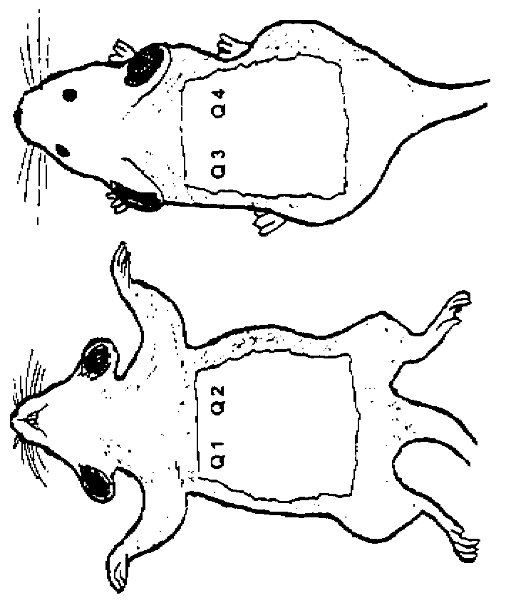
[0106] Intramuscular Implant Evaluation

[0107] To determine whether addition of DiCal would increase bone growth in implanted DBM-containing compositions, various test samples were implanted intramuscularly into athymic rats as described below. Surgery was performed on each rat as follows. Before surgery, rats were anesthetized by intraperitoneal injection of a ketamine / xylazine solution: 70 mg / kg of ketamine and 5 mg / kg of xylazine. General anesthesia was indicated by lack of response to toe pinch. Anesthesia was maintained with isoflurane if necessary.

[0108] The skin over the semitendinosus muscle was shaved (if necessary) using clippers and prepared by scrubbing with chlorhexadine and alcohol. Rats were placed in a lateral position. Using a scalpel or scissors, make a 1 cm incision in the skin along the long bone. Two 2 mm incisions were made in the semitendinosus muscle and blunt dissection was used to prepare the implantation bed.

[0109] Using aseptic techniq...
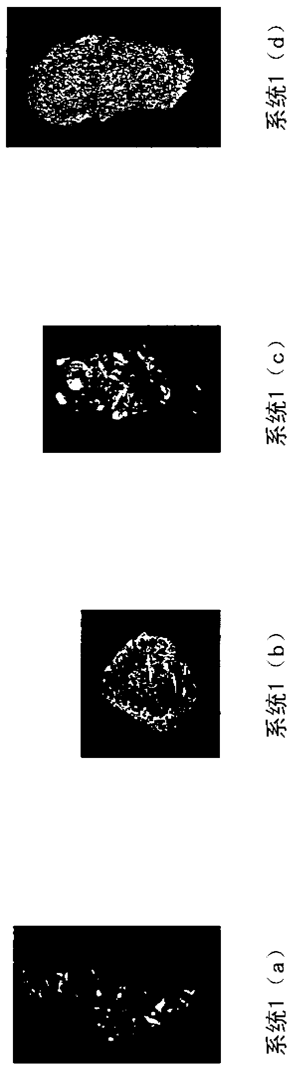
Embodiment 2

[0116] Subcutaneous Implant Evaluation

[0117] The various systems described below were implanted subcutaneously in athymic rats to evaluate the efficacy of the compositions on bone growth. Male athymic rats were used approximately 7 weeks of age at the start of the study. Surgery was performed on each rat as follows. Before surgery, rats were anesthetized by intraperitoneal injection of a ketamine / xylazine solution: 70 mg / kg of ketamine and 5 mg / kg of xylazine. General anesthesia was indicated by lack of response to toe pinch. Anesthesia was maintained with isoflurane if necessary.

[0118] The ventral and dorsal thorax regions of each rat were swabbed with chlorhexidine and alcohol swabs. Using a scalpel or scissors, make a 1 cm incision in the skin on the right side. This ventral incision is made at the base of the ribcage. After the incision, a pocket is made towards the axilla by blunt dissection under the skin and above the incision. Note the large blood vessels ...

Embodiment 3

[0125] Preparation of Exemplary Putty Compositions

[0126] In this embodiment, gelatin is combined with fibrous collagen, calcium and phosphate sources, and DMB microparticles to form a putty. By whipping 0.18g of collagen, 7.3mL of 30mM hydrochloric acid, 0.21g of calcium hydrogen phosphate (Dical) and 1.6g of demineralized bone microparticles into a slurry, and then freeze-drying the slurry into a 5cm×2cm×0.5cm sponge, The demineralized bone matrix sponge was thus prepared. A 12% w / v gelatin solution was prepared by adding 1.2 g of gelatin to 10 mL of water at about 37°C to about 50°C. Then, the prepared sponge is soaked in water for about 10 minutes to about 30 minutes. After absorbing the water, the sponge is compressed to remove excess water. Then, 1.9 mL of 12% gelatin (at about 37°C to about 50°C) was added to the sponge, spread using a spatula and mixed with the sponge. After 10 minutes, the mixture turned into a putty-like material. The putty material is cohesiv...


PUM


Abstract

A bone void filler composition containing an acidic mineral component that contains a calcium source and a phosphate source or a lower alkyl carboxylate source; an osteoinductive component that contains demineralized bone; and a three-dimensional, osteoconductive biologically acceptable carrier component that contains a collagenous material, and methods of making and using the composition, are described. The bone void filler composition may be in the form of a sponge of any desired shape or in the form of a paste or putty that is used to form a sponge or is obtained from particulated sponge, or a paste or putty that forms after a sponge is rehydrated. A pre-mixed bone void filler composition containing an acidic mineral component that contains a calcium source and a phosphate source or a lower alkyl carboxylate source; an osteoinductive component that contains demineralized bone; and a biologically acceptable carrier component that contains a liquid carrier, and methods of making and using the composition, are also described. The ratio of demineralized bone component to acidic mineral component may range from about 0.5:1 to about 80:1.

Description

[0001] Cross References to Related Applications [0002] This application is based on 35 U.S.C. § 119(e) claiming the benefit of U.S. Application No. 61 / 413,502, filed November 15, 2010, and U.S. Application No. 61 / 541,690, filed September 30, 2011, which Both applications are incorporated herein by reference. field of invention [0003] The field of the invention relates generally to bone void filler compositions, the preparation of said bone void filler compositions and the use of said bone void filler compositions, for example, to promote bone growth. Background technique [0004] In the fields of orthopedic surgery (eg, reconstruction, trauma, spine and dentistry), the effective repair of bone defects that may result from disease, injury, wound or surgery has long been a goal. Many materials and compositions have been considered, evaluated, or used. The biological, physical and mechanical properties of the materials and compositions become factors which, inter alia, af...

Claims


Application Information

Patent Timeline
IPC IPC(8): A61L24/00A61L27/46A61L27/36
CPCA61L24/0063A61L24/0084A61L27/3608A61L27/46A61L2300/414A61L2430/02A61P19/00C08L89/06
Inventor R·阿克拉I·R·加勒特H·B·文S·李D·M·阿克尔N·威尔海姆
Owner ZIMMER ORTHOBIOLOGICS
Why Eureka
Try Eureka