Preparation method of functionalized polyethyleneimine-modified multi-wall carbon nano-tube magnetic resonance imaging contrast agent
A technology of multi-walled carbon nanotubes and polyethyleneimine, which is applied in the field of preparation of multi-walled carbon nanotubes magnetic resonance imaging contrast agents, can solve problems such as long blood circulation time, improve blood pool imaging quality, and expand application range Effect
- Summary
- Abstract
- Description
- Claims
- Application Information
AI Technical Summary
Problems solved by technology
Method used
Image
Examples
Embodiment 1
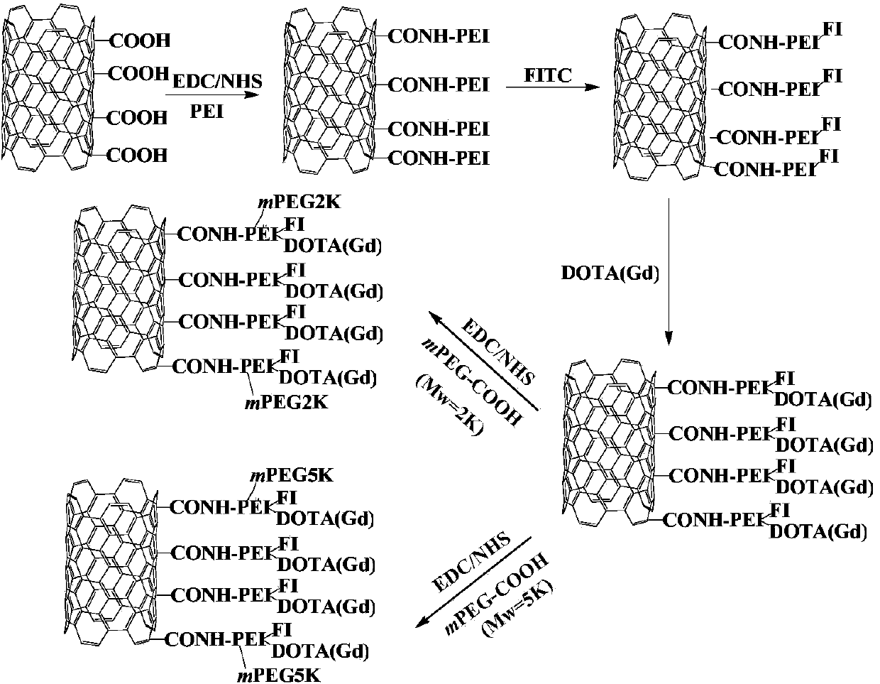
[0055] MWCNTs (100mg) were dispersed in 40mL of DMSO solution, and 10mL of DMSO solution containing EDC (120mg) was added with stirring, followed by 5mL of DMSO solution containing NHS (62mg), and the reaction was stirred at room temperature for 2h. Then 100 mg PEI (10 mL DMSO) was added dropwise to the DMSO solution of the EDC / NHS-activated MWCNTs, and the reaction was stirred for 2 days. Finally, put the reaction solution into a dialysis bag (MWCO=50,000), and remove the reaction solvent DMSO, excess reactants, and reaction by-products by dialysis, and dialyze three times with PBS buffer, 2L each time, and then dialyze with distilled water 5 times, 2L each time, and finally freeze-dry the aqueous solution of the product to obtain MWCNT / PEI;
[0056] Disperse 140 mg of prepared MWCNT / PEI in 50 mL of DMSO solution, add 8 mL of 4.85 mg of FI in DMSO solution dropwise under stirring, and stir at room temperature for 12 hours to obtain unpurified MWCNT / PEI-FI in DMSO solution; t...
Embodiment 2
[0064] T measured by in vitro MR 1 Value to test the MR imaging effect of the materials MWCNT / PEI-FI-DOTA(Gd) synthesized in Example 1, MWCNT / PEI-FI-DOTA(Gd)-mPEG2K and MWCNT / PEI-FI-DOTA(Gd)-mPEG5K. Take example 1 sample MWCNT / PEI-FI-DOTA (Gd) 8.46mg and dissolve it in 4.8mL of ultrapure water to prepare a solution with a gadolinium concentration of 0.892mM, then dilute it to a gadolinium concentration of 0.713, 0.535, 0.357, 0.178, 0.089 Each 1.5mL of the solution of mM; Get example 1 sample MWCNT / PEI-FI-DOTA (Gd)-mPEG2K11.52mg to be dissolved in the ultrapure water of 4.8mL and prepare the solution that gadolinium concentration is 0.675mM, then dilute to gadolinium concentration respectively 0.540, 0.405, 0.270, 0.135, each 1.5mL of 0.068mM solution; take example 1 sample MWCNT / PEI-FI-DOTA(Gd)-mPEG5K22.83mg and dissolve in 4.8mL of ultrapure water to prepare 0.876mM gadolinium concentration The solution was then diluted to 1.5mL each with gadolinium concentrations of 0.701,...
Embodiment 3
[0066] The cytotoxicity of the prepared material was investigated by MTT assay. Collect the logarithmic phase cells and add them to the 96-well cell culture plate, add 200 μL of cell-containing RPMI1640 medium to each well to make the cell density to 10000 / well, add 200 μL of sterile PBS buffer to the edge wells; then in the cell incubator (5 %CO 2 , 37°C) for 24 hours, until the cell monolayer covered the bottom of the well, discard the medium and add 180 μL of fresh medium, then add MWCNT / PEI-FI-DOTA(Gd), MWCNT / PEI-FI -20 μL PBS buffer solution of DOTA(Gd)-mPEG2K and MWCNT / PEI-FI-DOTA(Gd)-mPEG5K (each material is set to 7 concentrations based on gadolinium ion, namely 0, 2.5, 5, 10, 20, 40, 80μM) to verify the effect of the material itself on cell growth. All experimental groups were set up with 3 wells as a parallel group; after incubation in the incubator for 24 hours, 20 μL of MTT solution was added to each well, and the culture was continued for 4 hours to allow the ce...
PUM
 Abstract
Description
Claims
Application Information
 - R&D
- Intellectual Property
- Life Sciences
- Materials
- Tech Scout
- Unparalleled Data Quality
- Higher Quality Content
- 60% Fewer Hallucinations
Browse by: Latest US Patents, China's latest patents, Technical Efficacy Thesaurus, Application Domain, Technology Topic, Popular Technical Reports.
© 2025 PatSnap. All rights reserved.Legal|Privacy policy|Modern Slavery Act Transparency Statement|Sitemap|About US| Contact US: help@patsnap.com


