Method and culture medium for detecting sclerotinia sclerotiorum
A technology for Sclerotinia sclerotiorum and solid culture medium, which is applied in microorganism-based methods, chemical instruments and methods, biochemical equipment and methods, etc., can solve the problems of low specificity, low sensitivity, strong experience type, etc. High performance and high sensitivity
- Summary
- Abstract
- Description
- Claims
- Application Information
AI Technical Summary
Problems solved by technology
Method used
Image
Examples
Embodiment 1
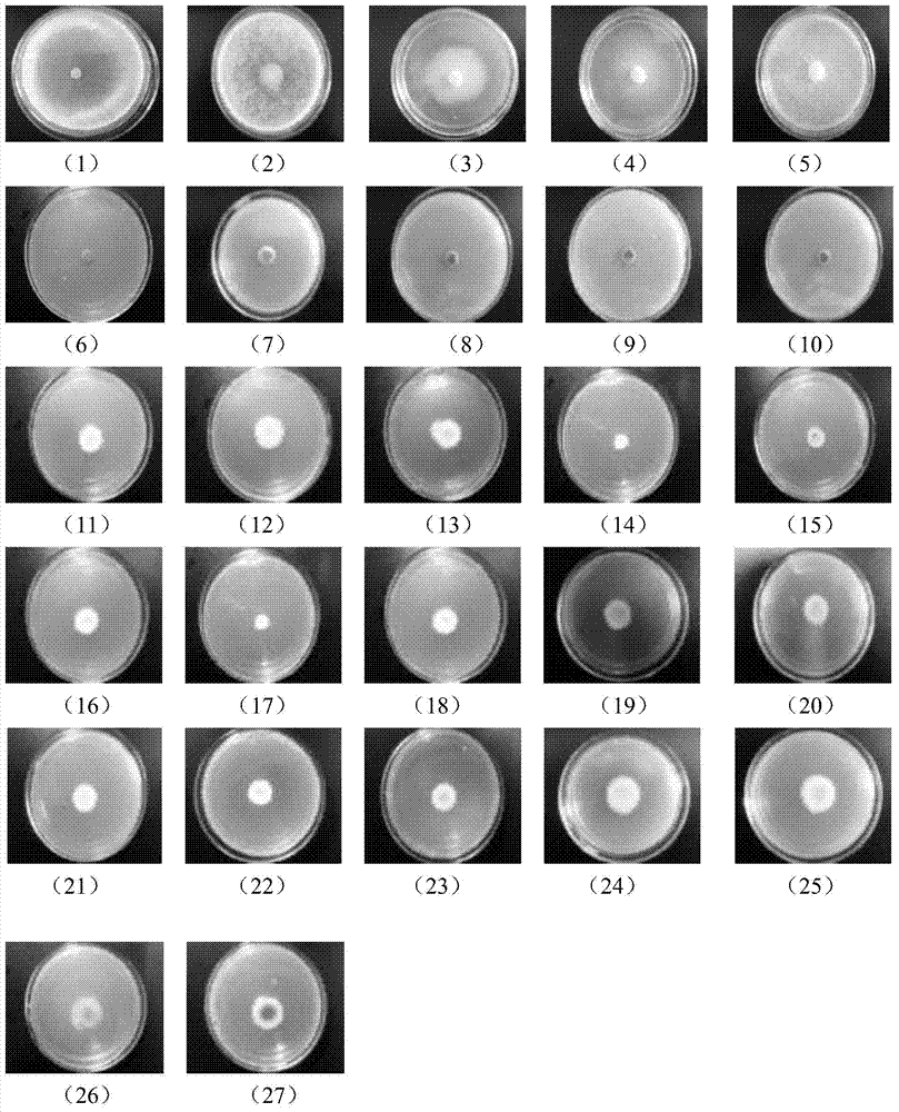
[0045] Five kinds of Sclerotinia sclerotiorum bacteria with different physiological characteristics were cultured on PDA medium to the logarithmic growth phase, and a 6 mm diameter plate was taken from the edge of the colony of the pathogenic bacteria to be tested, and inoculated in a medium containing no or containing 50 μg / mL detection Put the reagent on a PDA medium plate and culture it at a constant temperature of 25°C for 2 days. Observe the color of the colonies with the naked eye and the color of mycelia under an optical microscope. The results are as follows: figure 1 (1)~ figure 1 (10).
[0046] figure 1 (1)~ figure 1 (5) 5 species of Sclerotinia sclerotiorum Ss01, Hm25, Hm10 tested respectively I 、Hm10 T , and Hm10 IT When grown on a plate without detection reagents, the color of the colony is white, and the hyphae are colorless.
[0047] figure 1 (6-)~ figure 1 (10) 5 species of Sclerotinia sclerotiorum Ss01, Hm25, Hm10 tested respectively I 、Hm10 T , and ...
Embodiment 2
[0050] 17 kinds of different plant pathogenic bacteria such as Rhizoctonia solani, Sclerotinia watermelon, Botrytis cinerea, etc. were cultivated on PDA medium to logarithmic growth phase, and the diameter of 6mm bacterial discs were taken at the edge of the colony of the tested pathogens. Inoculate it on a PDA medium plate that does not contain or contain 50 μg / mL detection reagent, and culture at a constant temperature of 25°C for 2 days, observe the color of the colony with the naked eye, and observe the color of mycelia under an optical microscope. The results of culture with the detection reagent are as follows: figure 1 (11)~ figure 1 (27).
[0051] figure 1 (11)~ figure 1 (27) are the colony morphology and color of the 17 plant pathogenic bacteria tested. There was no difference in the color of the colonies of these pathogens grown on plates with and without the test reagent. Among them: Rhizoctonia solani, watermelon sclerotinia, mulberry sclerotinia, cabbage anthr...
Embodiment 3
[0053] Cultivate Sclerotinia sclerotiorum on PDA medium to the logarithmic growth phase, take a 6 mm diameter plate from the edge of the colony, inoculate it in a PSB shaker flask containing 50 μg / mL detection reagent, and place it at 25 ° C, 180 rpm Cultured at constant temperature for 3 days. Observe the color of mycelium and hyphae under a light microscope. The result is as figure 2 shown.
[0054] figure 2 (a) and (c) are the mycelium of Sclerotinia sclerotiorum cultured in the PSB shake flask without detection reagent, the mycelium is white, and the mycelium is colorless; figure 2 (b) and (d) are the mycelium of Sclerotinia sclerotiorum cultured in PSB shake flasks containing 50 μg / mL detection reagent, the mycelium is green, and the mycelium is green.
PUM
 Abstract
Description
Claims
Application Information
 - R&D
- Intellectual Property
- Life Sciences
- Materials
- Tech Scout
- Unparalleled Data Quality
- Higher Quality Content
- 60% Fewer Hallucinations
Browse by: Latest US Patents, China's latest patents, Technical Efficacy Thesaurus, Application Domain, Technology Topic, Popular Technical Reports.
© 2025 PatSnap. All rights reserved.Legal|Privacy policy|Modern Slavery Act Transparency Statement|Sitemap|About US| Contact US: help@patsnap.com


