Nanoparticle coupled with coupling cell-penetrating peptide and metal matrix proteinase (MMP) restriction enzyme digestion site

A technology of nanoparticles and enzyme cleavage sites, which is used in gene carriers, drug carriers, in vivo tissue cell imaging, and tumor treatment fields. control issues

Inactive Publication Date: 2014-05-14
TONGJI UNIV
View PDF4 Cites 17 Cited by
  • Summary
  • Abstract
  • Description
  • Claims
  • Application Information

AI Technical Summary

Problems solved by technology

However, although membrane-penetrating peptides have high-efficiency cell-penetrating ability, they lack tissue and cell specificity, and their application as a nanoparticle transport carrier is too complicated and difficult to control
The inefficiency of the target cells’ uptake of the simple penetrating peptide-mediated carrier and the problems of the absorption, distribution and bioavailability of the simple penetrating peptide-mediated carrier in the body seriously restrict its application as a delivery tool for nanoparticles

Method used


Image

  • Nanoparticle coupled with coupling cell-penetrating peptide and metal matrix proteinase (MMP) restriction enzyme digestion site
  • Nanoparticle coupled with coupling cell-penetrating peptide and metal matrix proteinase (MMP) restriction enzyme digestion site
  • Nanoparticle coupled with coupling cell-penetrating peptide and metal matrix proteinase (MMP) restriction enzyme digestion site

Examples


Embodiment 1

[0021] In vivo NMR imaging of magnetic nanospheres coupled with MAP-N membrane-penetrating peptide and MMP cleavage site

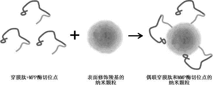
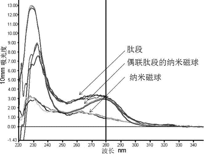
[0022] Solid-phase synthesis of KLKLALALALAPLGLAG peptide, which contains MAP-N penetrating peptide and MMP enzyme cleavage site sequence. By electrostatic self-assembly method, the surface of magnetic nanoparticles is sequentially modified with positively charged poly(diallyldimethylammonium chloride), PDADMAC and negatively charged polyacrylic acid (Poly(acrylic acid), PAA) to obtain carboxylated magnetic nanocomposite particles. Then, the carboxyl group on PAA was activated by 1-ethyl-3 (3-dimethylaminopropyl) carbodiimide hydrochloride (1-ethyl-3 (3-dimethylamino prowl)-carbodiimide, EDC) React with the amino group on the peptide (KLKLALALALAPLGLAG), so that the peptide is successfully connected to the surface of the magnetic particle ( figure 1 ).

[0023] Use the UV method at a wavelength of 280nm to directly test the peptide, and select the Warb...

Embodiment 2

[0025] Example 2: In vivo magnetothermal treatment of superparamagnetic nano-magnetic spheres coupled with SNS membrane-penetrating peptide and MMP cleavage site

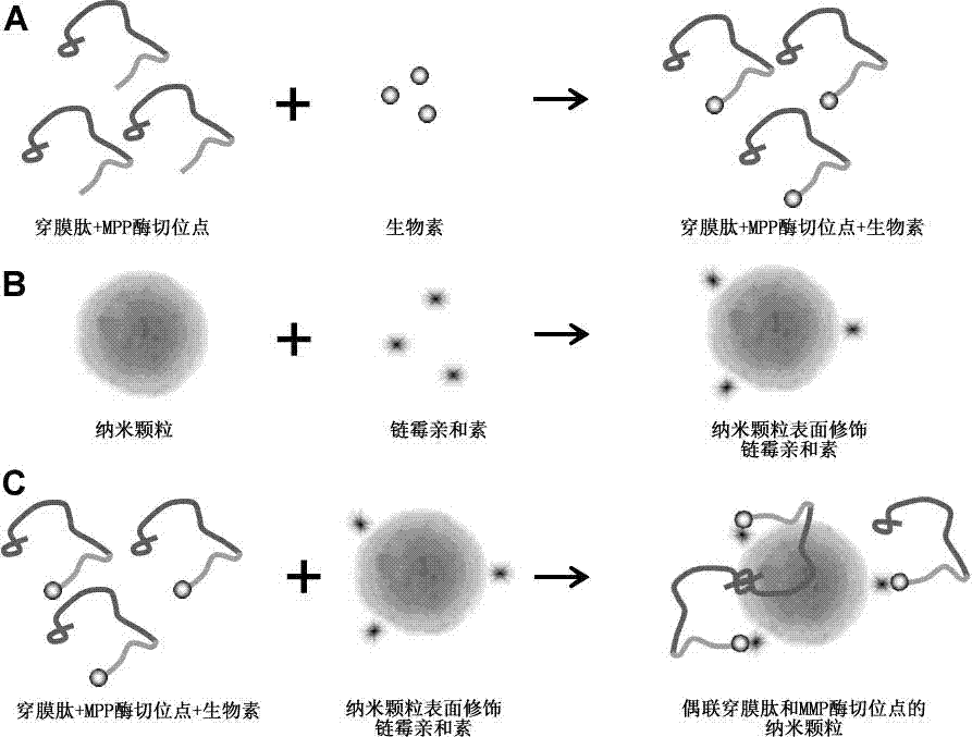
[0026] Solid-phase synthesis of AAVALLPAVLLALLAP PLGLAG peptide, which contains SNS penetrating peptide and MMP enzyme cleavage site sequence. AAVALLPAVLLALLAP PLGLAG peptide carboxyl-terminus linked to biotin. Surface modification of superparamagnetic nanospheres with streptavidin. Through the affinity reaction of biotin and streptavidin, the peptides were successfully linked to the surface of superparamagnetic nanospheres ( figure 2 ), the method for judging whether the peptide is coupled to the magnetic nanosphere is to directly test the peptide by UV method.

[0027] Inject superparamagnetic nanospheres coupled with peptides into the tail vein of the tumor-bearing animal model. After 24 hours, place the animal in an alternating magnetic field so that the internal temperature of the tumor site reaches 42°C and...


PUM


Abstract

The invention relates to a nanoparticle coupled with a coupling cell-penetrating peptide and a metal matrix proteinase (MMP) restriction enzyme digestion site. The cell-penetrating peptide and the MMP restriction enzyme digestion site are distributed outside the surface-modified carboxyl or streptavidin nanoparticle; the cell-penetrating peptide and the MMP restriction enzyme digestion site are ligated via a chemical bond, and the amino group of the last amino acid residue of the restriction enzyme digestion site sequence is ligated with the carboxyl group on the surface of the nanoparticle by condensation reaction; or the last amino acid residue of the restriction enzyme digestion site sequence is ligated with biotin, the surface of the nanoparticle is modified with streptavidin, and a peptide fragment is ligated with the nanoparticle by use of the biotin-streptavidin affinity reaction. According to the invention, an MMP restriction enzyme digested peptide segment is innovatively utilized to couple the coupling cell-penetrating peptide and the nanoparticle, and the cell-penetrating peptide is separated from the nanoparticle by the fact that the activity of MMP inside or around a target cell is higher than the activity of MMP inside or around a normal cell, and physical or drug therapy, imaging and other treatment are performed on the target cell and tissue by use of the specific physiological environment inside or around the target cell or the surrounding auxiliary physical environment and the characteristics of the nanoparticle.

Description

technical field [0001] The invention belongs to the fields of tumor treatment, drug carrier, live tissue cell imaging and gene carrier, and specifically relates to a nanoparticle coupled with a membrane-penetrating peptide and an MMP enzyme cleavage site. Background technique [0002] At present, more and more nanoparticles (superparamagnetic magnetic spheres, molecular polymers, gold particles, etc.) have been found to have very good drug loading, imaging and gene carrier functions. The vast majority of nanoparticles cannot enter cells through active diffusion or passive transport due to their diameter, surface charge, polarity distribution, and the biofilm barrier effect of the cell itself. The active uptake of nanoparticles by target cells in vivo is very small. Therefore, the lack of ability to penetrate cell membranes and reach intracellular targets is an important obstacle in the research and development of nanoparticles. [0003] Nanoparticles that are difficult to ...

Claims


Application Information

Patent Timeline
Patent Type & Authority Applications(China)
IPC IPC(8): A61K9/14A61K47/42A61K41/00A61K49/00A61P35/00
Inventor 李卓权林耀忠
Owner TONGJI UNIV
Why Eureka
Try Eureka