
Endocervical cell preserving fluid and method for preparing endocervical cell specimen

A cervical cell and preservation solution technology, which is applied to animal cells, vertebrate cells, artificial cell constructs, etc., can solve the problems of easy fatigue of examiners when reading images, not suitable for cervical cancer screening, and easy to contaminate transportation personnel and tools. , to achieve the effect of clear and clean smear background, time-saving and labor-saving observation, and easy popularization and promotion.

Inactive Publication Date: 2015-03-18
THE MILITARY GENERAL HOSPITAL OF BEIJING PLA
View PDF3 Cites 0 Cited by
  • Summary
  • Abstract
  • Description
  • Claims
  • Application Information

AI Technical Summary

Problems solved by technology

The traditional Pap direct smear method is the main method for early screening of cervical cancer and has been used for more than 50 years. However, the traditional Pap direct smear method has the following disadvantages: the sample collection uses cotton swabs or cork chips, which is easy to contaminate transport personnel and tools; only 20% of the cells collected by direct smear are applied on the glass slide, and there are too few diagnostic components of the cells; Excessive force during filming will deform the cells; it is easy to dry the film, causing cell rupture and making it difficult to diagnose; discarding the sampler, about 80% of the cell samples are left on the sampler and discarded, and abnormal cells that may be found are also discarded; Cell specimens cannot be preserved and cannot be made into slides; a large amount of mucus, red blood cells, and white blood cells cover the smear, resulting in unclear background and poor staining of the smear, which is not conducive to diagnosis and affects the detection rate to a certain extent
However, the "cervical cell preservation solution" used in this technology is mostly imported from abroad, the price is very expensive, the source is inconvenient, and it cannot be popularized, so it cannot be used for extensive cervical cancer screening.
The cytocentrifuge or membrane-type ultra-thin cell slicer in this technology is also very expensive, and some small and medium-sized hospitals cannot afford it.

Method used


Image

  • Endocervical cell preserving fluid and method for preparing endocervical cell specimen
  • Endocervical cell preserving fluid and method for preparing endocervical cell specimen
  • Endocervical cell preserving fluid and method for preparing endocervical cell specimen

Examples


Embodiment 1

[0053] 1 Materials and methods

[0054] 1.1 Source of samples: A total of 1,450 gynecological specimens from the outpatient department of Beijing Military Region General Hospital for cervical cancer screening were selected from January 2012 to January 2013. Traditional Pap smears and liquid-based cytological preparations were performed respectively. The detection rate of positive lesions and the quality of film production by different production methods.

[0055] 1.2 Preparation of cervical cell preservation solution

[0056] Weigh 9g of N-acetyl-3-mercaptoalanine, 34g of sodium chloride, 14g of disodium hydrogen phosphate, 1g of sodium dihydrogen phosphate, measure 4000mL of absolute alcohol, 48mL of glacial acetic acid, 5mL of concentrated hydrochloric acid, and 4000M1 of distilled water, Use after dissolving.

[0057] 1.3 Instruments Oscillator, centrifuge and smear device.

[0058] 1.4 Sample collection and production methods

[0059] 1.41 Traditional Pap smear method:...

Embodiment 2

[0078] The preparation of cervical cell preservation solution in the present embodiment two:

[0079] Weigh 5g of N-acetyl-3-mercaptoalanine, 30g of sodium chloride, 10g of disodium hydrogen phosphate and 0.7g of sodium dihydrogen phosphate and dissolve them in a small amount of water, then measure 4500mL of absolute alcohol, 40mL of glacial acetic acid and concentrated 4mL of hydrochloric acid, and finally dilute to 10000mL for later use.

[0080] Use the cervical cell preservation solution of the above formula to prepare cervical cell specimens according to the following method:

[0081] Step 1: Under the direct vision of the vaginal dilator, the gynecologist wipes off the secretions on the surface of the cervix and vagina with a cotton swab, then inserts the cervical brush into the cervical canal along the external os of the cervix, rotates clockwise 3 times, and takes out the cervical brush. Soak the brush head in the collection tube filled with cervical cell preservation...

Embodiment 3

[0091] Weigh 15g of N-acetyl-3-mercaptoalanine, 50g of sodium chloride, 20g of disodium hydrogen phosphate and 1.4g of sodium dihydrogen phosphate and dissolve them in a small amount of water, then measure 5500mL of absolute alcohol, 80mL of glacial acetic acid and concentrated Hydrochloric acid 8mL, and finally dilute to 10000mL to make cervical cell preservation solution for later use.

[0092] Use the cervical cell preservation solution of the above formula to prepare cervical cell specimens according to the following method:

[0093] Step 1. Under the direct vision of the vaginal dilator, the gynecologist wipes off the secretions on the surface of the cervix and vagina with a cotton swab, then inserts the cervical brush into the cervical canal along the external opening of the cervix, rotates clockwise 5 times, and takes out the cervical brush. Soak the brush head in the collection tube filled with cervical cell preservation solution, tighten the cap of the tube, label it,...


PUM


Abstract

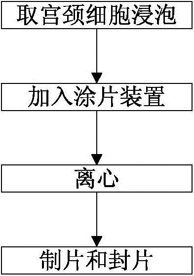
The invention discloses endocervical cell preserving fluid and a method for preparing an endocervical cell specimen. The endocervical cell preserving fluid comprises N-acetyl-3-cysteine, anhydrous alcohol, sodium chloride, disodium hydrogen phosphate, sodium dihydrogen phosphate, glacial acetic acid, concentrated hydrochloric acid and distilled water. The method for preparing the endocervical cell specimen comprises the following steps; soaking endocervical cells in a collection tube of the endocervical cell preserving fluid, uniformly oscillating, adding the endocervical cells in a smear device, arranging the smear device in a centrifugal machine for centrifuging, and taking a glass slide with a uniform thin-layer cell smear; and fixing the obtained glass slide with the cell smear by a fixing solution, dyeing, dehydrating, making the glass slide transparent, and finally, mounting to obtain a specimen smear for diagnosis. The endocervical cell preserving fluid and the method for preparing the endocervical cell specimen are reliable and practical, are low in cost, and have useful effects, thus being suitable for large-scale popularization.

Description

technical field [0001] The invention relates to a cervical cell preservation solution and a preparation method of cervical cell specimens. Background technique [0002] Cervical cancer is one of the most common gynecological malignancies, so the screening of cervical cancer has been widely concerned. The traditional Pap direct smear method is the main method for early screening of cervical cancer and has been used for more than 50 years. However, the traditional Pap direct smear method has the following disadvantages: the sample collection uses cotton swabs or cork chips, which is easy to contaminate transport personnel and tools; only 20% of the cells collected by direct smear are applied on the glass slide, and there are too few diagnostic components of the cells; Excessive force during filming will deform the cells; it is easy to dry the film, causing cell rupture and making it difficult to diagnose; discarding the sampler, about 80% of the cell samples are left on the s...

Claims


Application Information

Patent Timeline
Patent Type & Authority Patents(China)
IPC IPC(8): C12N5/071A01N1/00G01N1/28
Inventor 田玉旺
Owner THE MILITARY GENERAL HOSPITAL OF BEIJING PLA
Learn More