
A method for observing the microtubule structure of the cytoskeleton in animal liver tissue

A technology of animal liver and cytoskeleton, applied in biological testing, preparation of test samples, fluorescence/phosphorescence, etc., to achieve the effect of easy mastery, strong practicability and scalability, and wide application range

Active Publication Date: 2016-06-15
NANJING INST OF ENVIRONMENTAL SCI MINIST OF ECOLOGY & ENVIRONMENT OF THE PEOPLES REPUBLIC OF CHINA
View PDF5 Cites 0 Cited by
  • Summary
  • Abstract
  • Description
  • Claims
  • Application Information

AI Technical Summary

Problems solved by technology

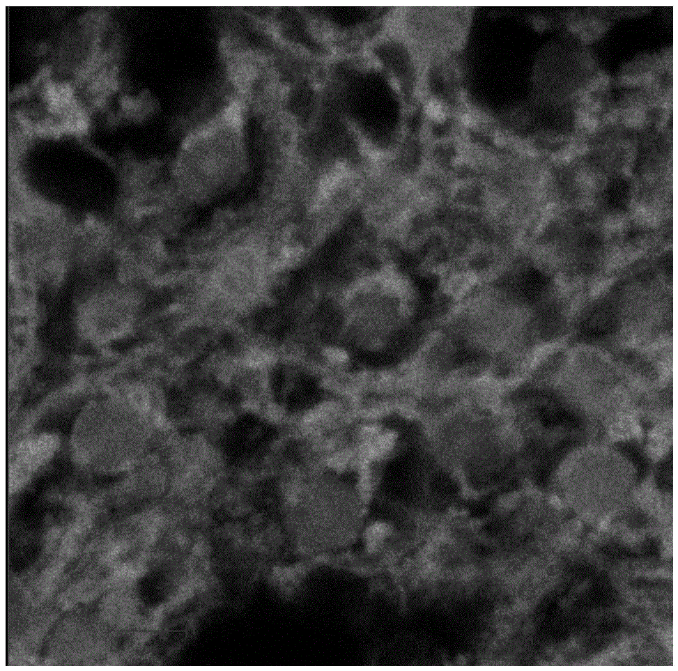
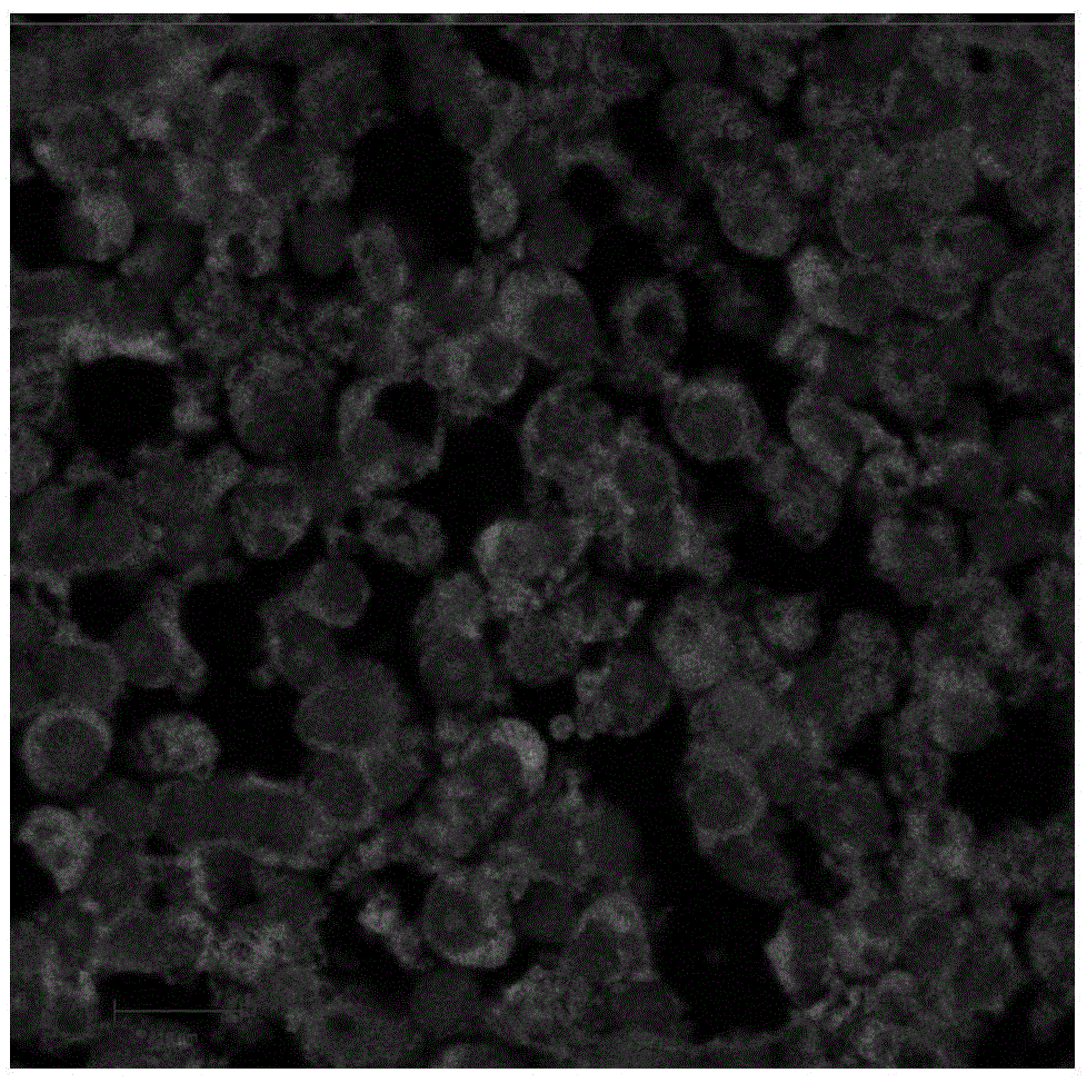
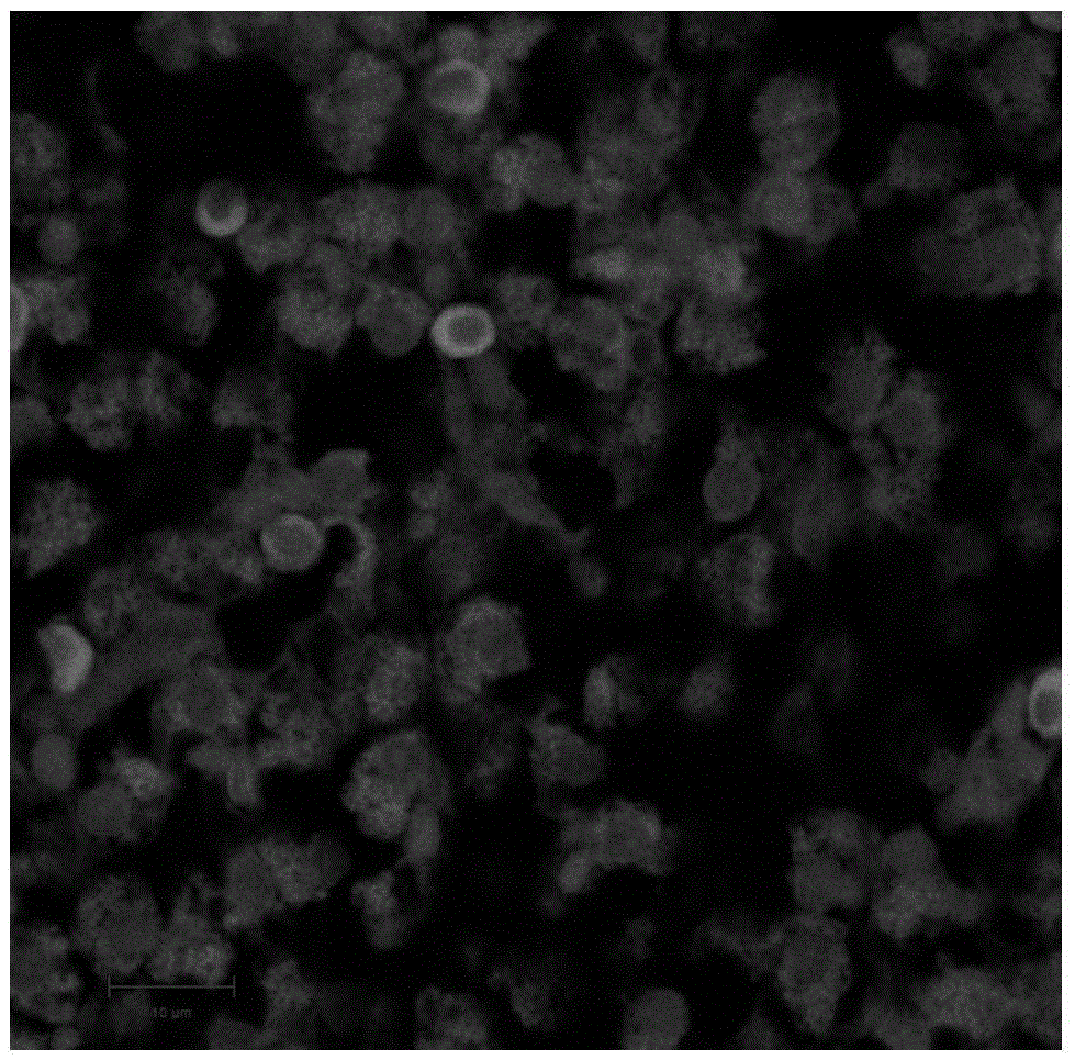
[0005] The technical problem to be solved by the present invention is to provide a method for observing the microtubule structure of the cytoskeleton in the animal liver tissue for the deficiency of the existing cytoskeleton system observation method, using β-Tubulin (9F3) Rabbit (Alexa 488Conjugate) monoclonal antibody (Sigma) was used as the primary antibody, combined with tubulin in the cytoskeleton in animal liver slices, and laser scanning confocal technology was used to observe the cytoskeleton structure, which can achieve accurate and intuitive observation of the cytoskeleton system and prevent pollution Provide direct evidence for the reorganization of animal liver tissue cytoskeleton system caused by chemical stress

Method used


Image

  • A method for observing the microtubule structure of the cytoskeleton in animal liver tissue
  • A method for observing the microtubule structure of the cytoskeleton in animal liver tissue
  • A method for observing the microtubule structure of the cytoskeleton in animal liver tissue

Examples


Embodiment 1

[0032] Example 1: Imaging of normal liver cytoskeleton system of carp (Cyprinus carpio) by laser scanning confocal microscope.

[0033] Young (about 6 months) carps purchased from a fishery in the Freshwater Fishery Research Center of the Chinese Academy of Fishery Sciences were selected as the test organisms, with an average body length and weight of 14.00±1.08cm and 29.26±5.09g, respectively. Domesticated in aerated tap water for 14 days. During domestication, the environmental conditions were as follows: water temperature (16.1±0.2°C), pH value (7.20±0.35), dissolved oxygen (8.6±0.5mg / L), light-dark ratio (12h / 12h) , converted to CaCO 3 total hardness (129.7±8.3mg / L). Maintain aeration during acclimatization and experiments. Feeding was withheld 2 days before exposure and fed once a day during the two-day exposure period.

[0034] After domestication, several healthy fish were randomly selected and sacrificed on ice, and the fish livers were quickly taken out, washed wit...

Embodiment 2

[0036] Example 2: Changes in the cytoskeleton system of carp (Cyprinus carpio) liver under microcystin stress.

[0037] Young (about 6 months) carps purchased from a fishery in the Freshwater Fishery Research Center of the Chinese Academy of Fishery Sciences were selected as the test organisms, with an average body length and weight of 14.00±1.08cm and 29.26±5.09g, respectively. Before the experiment, carp were domesticated in aerated tap water for 14 days, and they were randomly divided into two groups, and each group was subdivided into two groups according to the exposure time, with 6 fish in each treatment group. The two groups of fish were intraperitoneally injected with sublethal doses of 50 μg / kg and 120 μg / kg (MC-LR was dissolved in normal saline) respectively according to MC-LR, and then were sacrificed on ice after 5 and 12 hours respectively, and the fish liver was quickly taken out, according to Example 1 According to the method, the specimen is pre-treated, and af...

Embodiment 3

[0040] Example 3: Effects of the joint action of microcystin and butythionimide on the cytoskeleton system of carp (Cyprinus carpio) liver.

[0041] Young (about 6 months) carps purchased from a fishery in the Freshwater Fishery Research Center of the Chinese Academy of Fishery Sciences were selected as the test organisms, with an average body length and weight of 14.00±1.08cm and 29.26±5.09g, respectively. Before the experiment, carp were domesticated in aerated tap water for 14 days, and were randomly divided into three groups, with 6 fish in each treatment group. Two intraperitoneal injections of sublethal doses of 50 μg / kg MC-LR (dissolved in normal saline), 200 mg / kg BSO, and 200 mg / kg BSO+50 μg / kg MC-LR (pre-injection of BSO200 mg / kg 1 h before intraperitoneal injection of 50 μg / kg MC-LR), respectively After acting for 48 hours, the fish was put to death on ice, and the cod liver was quickly taken out, and the specimen was pretreated according to the method described in ...


PUM

PropertyMeasurementUnit
thicknessaaaaaaaaaa
diameteraaaaaaaaaa
hardnessaaaaaaaaaa

Abstract

The invention discloses a method for observing a microtubule structure of a cytoskeleton in a liver tissue of animals. The method comprises the following steps: (1) cleaning the liver tissue of animals with PBS, fixing for 8-12h with 4v / v% of paraformaldehyde water solution, and carrying out paraffin embedding and serial sectioning; (2) dewaxing the obtained paraffin sections, and carrying out antigen retrieval, closing and fluorescently labeled antibody incubation; (3) for specimens sealed by glycerol, observing through a laser scanning confocal microscope within 24h, respectively exciting 488 and 4' 6-diamidino-2-phenylindole under 488nm and 405nm, and observing and shooting at once at room temperature in a dark room environment. The method is fast, simple, intuitive and accurate, is strong in practicality and extensibility, and can research the dose-effect changing relationship of the cytoskeleton under stress of pollutants.

Description

technical field [0001] The invention relates to a method for observing changes in the cytoskeleton system of animal liver tissue by using a laser scanning confocal microscope, more specifically a method for observing the expression of tubulin (β-tubulin) in fish liver tissue slices by using a laser scanning confocal microscope and distribution methods. Background technique [0002] Cytoskeleton refers to the protein fiber network structure in eukaryotic cells, through which eukaryotic cells can maintain their basic shape, so this important structure is vividly called cytoskeleton, and it is usually considered to be an organelle in a broad sense kind of. The cytoskeleton not only plays an important role in maintaining cell shape, bearing external forces, and maintaining the orderliness of the internal structure of the cell, but also participates in many important life activities, such as: cytoskeleton traction chromosome separation in cell division, and cell material transpo...

Claims


Application Information

Patent Timeline
Patent Type & Authority Patents(China)
IPC IPC(8): G01N33/68G01N21/64
CPCG01N1/28G01N21/6402G01N33/68
Inventor 姜锦林单正军卜元卿程燕王晓蓉
Owner NANJING INST OF ENVIRONMENTAL SCI MINIST OF ECOLOGY & ENVIRONMENT OF THE PEOPLES REPUBLIC OF CHINA
Learn More