

Method for detecting hemoglobin-combined alpha-synuclein
A technology for synuclein and hemoglobin, which is applied in the field of detecting the content of α-synuclein bound to hemoglobin in blood red blood cells, can solve the problems of inconclusive diagnosis of Parkinson's disease and the like
- Summary
- Abstract
- Description
- Claims
- Application Information
AI Technical Summary
Problems solved by technology
Method used
Image


Examples
Embodiment 1
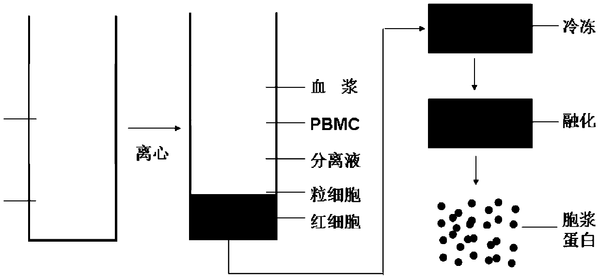
[0119] Preparation of erythrocyte cytoplasmic protein ( figure 1 )
[0120] step 1,
[0121] Take 5-10ml anticoagulant blood (EDTA, heparin or citrate anticoagulant). Gently mix the anticoagulated blood upside down, add it into a 50ml centrifuge tube, record the volume of whole blood, add PBS at a ratio of 1:1, and mix well. Step 2,
[0122] Slowly add the diluted whole blood to the lymphocyte separation medium, centrifuge at 400×g for 20min. At this time, the order of the liquid in the tube from top to bottom is the plasma layer, the buffy coat layer (peripheral blood mononuclear cells), the separation liquid layer and the red blood cell layer.
[0123] Step 3,
[0124] Aspirate off the plasma layer, buffy coat layer and separation liquid layer, transfer the red blood cells at the bottom layer to a new 50ml centrifuge tube, add PBS to 40ml, centrifuge at 2000rpm for 10min, and repeat this 3 times. Aliquot and store at -80°C for later use.
[0125] Step 4,
[0126] Fr...
Embodiment 2
[0128] ELISA detection process of hemoglobin-binding α-synuclein
[0129] step 1,
[0130] Coating: with NaHCO 3 The buffer solution dilutes the anti-human hemoglobin antibody to a final concentration of about 0.1-2 μg / mL. Add 100 μL of the antibody dilution to each well of the microtiter plate and incubate overnight at 4°C. Rinse each well of the microtiter plate with PBST (which is PBS containing Tween), and determine the number and time of washing as required.
[0131] Step 2,
[0132] Blocking: Add 100 μL of blocking solution (PBS containing gelatin and Tween-20) to each well of the microtiter plate, and incubate at 37° C. for 1 to 2 hours. Rinse each well of the microtiter plate with PBST, and determine the number and time of washing as needed.
[0133] Step 3,
[0134] Adding samples: Add 100 μL of the prepared red blood cell cytoplasm sample to each well at a concentration of 8-10 μg / mL, and incubate at 37°C for 1-2 hours. Rinse each well of the microtiter plate...
Embodiment 3
[0144] Detection of real samples ( figure 2 )
[0145] The relative quantitative ELISA method was used to detect the amount of α-synuclein combined with hemoglobin in the blood erythrocyte cytoplasm of 100 clinically diagnosed Parkinson's patients and 100 healthy controls. The results showed that the amount of α-synuclein bound to hemoglobin in the erythrocytes of Parkinson's patients was 0.83 μmol / L, and the highest value of α-synuclein bound to hemoglobin in the erythrocytes of Parkinson's patients was 1.025 μmol / L; The amount of α-synuclein bound to hemoglobin in normal healthy control red blood cells was 0.454 μmol / L, and the highest value was 0.597 μmol / L. The amount of α-synuclein combined with hemoglobin in the red blood cells of Parson's patients was significantly higher than that of normal healthy controls (p<0.01).
PUM
 Abstract
Description
Claims
Application Information