
Nanometer probe for detecting activity of intracellular telomerase in situ and preparation method thereof

A nano-probe, in-situ detection technology, applied in biochemical equipment and methods, measurement devices, microbial determination/inspection, etc. Effect

Active Publication Date: 2014-12-31
NANJING UNIV
View PDF1 Cites 6 Cited by
  • Summary
  • Abstract
  • Description
  • Claims
  • Application Information

AI Technical Summary

Problems solved by technology

These technologies include telomerase purification method, optical detection method, array and biosensor chip, electrochemical analysis method, and biological barcoding technology, all of which require pre-extraction of cell fluid and cannot directly detect telomerase activity in living cells in situ

Method used


Image

  • Nanometer probe for detecting activity of intracellular telomerase in situ and preparation method thereof
  • Nanometer probe for detecting activity of intracellular telomerase in situ and preparation method thereof

Examples


Embodiment 1

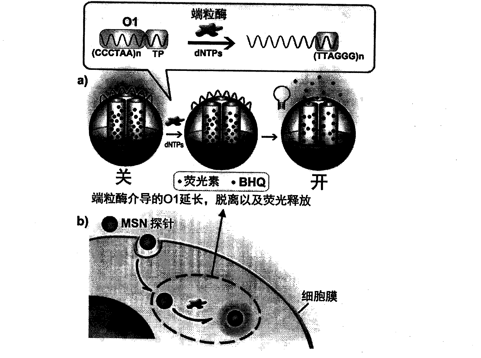
[0018] Example 1: Combining figure 1 , Synthesis of nanoprobes for in situ detection of intracellular telomerase activity

[0019] Add 1 mg of carbodiimide (EDC), 2.5 mg of N-hydroxysuccinimide (NHS) and 0.1 mg of carboxyl BHQ to 1 mL of 1 mg / mL aminated MSN dispersion, and stir magnetically for 4 hours at room temperature. MSN-BHQ was obtained after centrifugation and washing. Disperse MSN-BHQ in 1 mL of water and add 0.1 mg of fluorescein, and keep stirring overnight at room temperature. Centrifuge and wash the precipitate with ultrapure water and dry to obtain fluorescein MSN-BHQ. Disperse 1 mg of fluorescein MSN-BHQ in 1 mL of hybridization buffer (containing 10 mM Tris hydrochloride, 1 mM EDTA, 50 mM NaCl and 10 mM MgCl 2 ), add 10 μL (100 μM) O1, continue stirring at 37° C. for 1 hour, and centrifuge to obtain the MSN probe. Probes were dispersed in 1mL HB and stored at 4°C.

Embodiment 2

[0020] Example 2: Combining figure 2 , In situ detection of intracellular telomerase activity using nanoprobes

[0021] Using HeLa cervical cancer cells as a model, HeLa cells (0.5 mL, 1×10 6 mL -1 ) were cultured in 20-mm confocal dishes at 37°C for 24 hours. Then, 15 μL of MSN probe (1 mg / mL) was added to the culture dish, incubated at 37° C. for 1.5 hours, and then detected with a laser confocal fluorescence microscope. In telomerase-positive HeLa cells, the pore channel of the MSN probe was opened to release fluorescein, and a fluorescent signal appeared in the cytoplasm, realizing in situ imaging of telomerase activity in the cell. Cells (0.5 mL, 1×10 6 mL -1 ) were co-cultured for 48 hours, then 15 μL of MSN probe (1 mg / mL) was added to the petri dish, and after incubation at 37° C. for 1.5 hours, the change of telomerase activity after drug action was observed under a confocal microscope.


PUM


Abstract

The invention relates to a nanometer probe for detecting activity of intracellular telomerase in situ and a preparation method thereof. The prepared nanometer probe employs multihole silicon nanoparticle (MSN) as a carrier, the pore channel of the nanoparticle is covalently fixedly provided with a fluorescence quencher BHQ and is filled with fluorescein, and the surface of the nanoparticle is wrapped with DNA chain O1 of telomerase substrate fragment (TP) through electrostatic adsorption, so that fluorescein is plugged in the pore channel. BHQ quenches fluorescence of fluorescein, and the fluorescence state of the nanometer probe is off. Under the effect of telomerase, the 3' end of O1 on the surface of the probe is extended, and a plurality of TTAGGG fragments complementing 5' end are generated, so that the two ends hybridize and a cyclic structure is formed and breaks away from the probe surface, and thus the pore channel is opened and fluorescein in the probe is released, and the fluorescence state is transferred into open. The probe and cells are incubated, the probe enters cytoplasm, the probe pore channel is opened under the effect of telomerase in cytoplasm, and fluorescence generated by released fluorescein is utilized to realize in-situ detection on activity of intracellular telomerase.

Description

1. Technical field [0001] The invention relates to a nanometer probe for in situ detection of intracellular telomerase activity and a preparation method thereof. 2. Background technology [0002] Telomerase is a ribonucleoprotein complex composed of RNA and protein, and is a type of reverse transcriptase. Telomerase uses its internal RNA as a template to carry out reverse transcription in cells, making telomeres increase TTAGGG repeat sequence to maintain telomere length. In normal human cells, the activity of telomerase is quite tightly regulated and can only be detected in cells that must constantly divide, such as hematopoietic cells, stem cells and germ cells. In tumor cells, the activity of telomerase is activated, so it is an important basis for diagnosis and prediction of cancer. [0003] Currently, the most widely used assay for telomerase activity is the Telomere Repeat Amplification Program (TRAP). TRAP uses artificially synthesized sequences without telomeres a...

Claims


Application Information

Patent Timeline
IPC IPC(8): C12Q1/68C12Q1/48G01N21/64
CPCC12Q1/485G01N2333/9128
Inventor 丁霖钱若灿鞠熀先
Owner NANJING UNIV
Learn More