Medical multi-functional hemostasis material and preparation method thereof

A hemostatic material and multifunctional technology, applied in medical science, bandages, absorbent pads, etc., can solve the problems of mixed materials such as unsatisfactory toughness, poor blood concentration effect, unsatisfactory hemostasis efficiency, etc., and achieve wide clinical application value, Enhance the hemostatic effect and prevent adhesion

Inactive Publication Date: 2015-01-07
BEIJING JISHUITAN HOSPITAL
View PDF6 Cites 3 Cited by
  • Summary
  • Abstract
  • Description
  • Claims
  • Application Information

AI Technical Summary

Problems solved by technology

However, the water absorption capacity of a single collagen hemostatic material is limited, and the blood concentration effect is not good, so the hemostatic efficiency is not ideal
Moreover, collagen with a single structure is prone to adhesion in the body after sucking blood.
At present, the hemostatic materials used in the market have dev

Method used


Image

  • Medical multi-functional hemostasis material and preparation method thereof

Examples


preparation example Construction

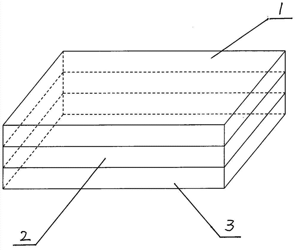
[0021] A. Preparation of hemostatic layer:

[0022] Add 0.2-2 parts of collagen, 0.1-0.6 parts of calcium chloride and 0.5-1 part of glycerin to 50-100 parts of 3% acetic acid solution, stir well, pour into a negative pressure tank, and discharge the air bubbles , to obtain a hemostatic collagen sponge solution; add 0.1-20 parts of polymer to 100 parts of an organic solvent to obtain a polymer organic solution, and then emulsify the hemostatic collagen sponge solution with a homogenizer and cast a film, and obtain after the organic solvent volatilizes The hemostatic layer solution is ready for use; or add 0.1-5 parts of sodium chloride to the polymer organic solution, cast the film, and after the organic solvent volatilizes, remove the sodium chloride particles in the polymer film by soaking, and then compound with the hemostatic collagen sponge solution Obtain the hemostatic layer solution for use.

[0023] B. Preparation of barrier layer:

[0024] Add 0.2-2 parts of collag...

Embodiment 1

[0029] Embodiment 1, the technology that adopts the original tissue of animal to prepare collagen used in the present invention is as follows:

[0030] (1) Remove the superficial fascia from the washed Achilles tendon, shred it, and air-dry it. 5 grams of air-dried bovine Achilles tendon fragments were placed in a beaker, and 500 mL of 3% acetic acid solution was added.

[0031] (2) Swell the solution prepared in step (1) for 24 hours at 4°C. Add 2.5 grams of pepsin to the solution, and stir with a homogenizer for 20 minutes. Seal the beaker and perform enzymatic hydrolysis for 72 hours at 4°C.

[0032] (3) Centrifuge the solution prepared in step (2) at 2500g for 20 minutes at 4°C. At room temperature, collect the supernatant after centrifugation.

[0033] (4) At normal temperature, add an equal volume of saturated sodium chloride solution to the supernatant collected in step (3), salt out the precipitate, centrifuge at 2500 g for 20 minutes, and collect the precipitate. ...

Embodiment 2

[0036] Embodiment 2, adopt commercially available collagen primary product to prepare the technique of collagen used in the present invention as follows:

[0037] Take 5 grams of commercially available collagen primary products, put them in a beaker, add 500 mL of 3% acetic acid solution, and then add 2.5 grams of pepsin, stir in a homogenizer for 20 minutes, seal the beaker, and enzymolyze 72 hours to produce collagen. Subsequent preparation is carried out according to the above-mentioned process steps for preparing the medical multifunctional hemostatic material.


PUM

PropertyMeasurementUnit
Thicknessaaaaaaaaaa
Thicknessaaaaaaaaaa
Thicknessaaaaaaaaaa

Abstract

The invention discloses a medical multi-functional hemostasis material in a three-layer structure formed by combining a hemostasis layer, a separation layer and an anti-adhesion layer by a casting method. The hemostasis layer which is a first layer is prepared by mixing calcium chloride, glycerine, collagen, an acetic acid solution, polymers or/and sodium chloride; the separation layer which is a middle layer is prepared by mixing the collagen and the acetic acid solution; the anti-adhesion layer which is a third layer is prepared by mixing sodium hyaluronate, 3% of an acetic acid solution, the polymer or/and the sodium chloride; the hemostasis layer, the separation layer and the anti-adhesion layer are combined into the three-layer structure in sequence by the casting method, and the medical multi-functional hemostasis material is formed through packaging and sterilization. Through the adoption of the medical multi-functional hemostasis material disclosed by the invention, the water absorption and the tenacity of the material are improved, the degradation of the material in bodies is delayed, the hemostasis effect is enhanced, the adhesion is prevented, and the material has the function of promoting tissue repair, in addition, the production process is simple, the making price of products is lower, and the material has large clinical popularization and application values.

Description

Technical field [0001] The invention relates to the field of medical biological materials, in particular to a medical multifunctional hemostatic material and a preparation method thereof. Background technique [0002] As we all know, bleeding is a common phenomenon in surgical operations, wound injuries and various accidents, which will bring many risks and harms to people's health and life. At the same time, the fibrin exuded from the blood during bleeding causes the deposition of fibrous tissue and protein, which is prone to adhesion at the bleeding site, which is not conducive to wound healing. And heavy hemorrhage directly threatens human or animal life. Surveys show that more than half of trauma patients die from hemorrhage. Therefore, fast, ideal and effective hemostatic and anti-adhesion materials and methods have always been the focus of research at home and abroad. Materials scientists and medical scientists in various countries are trying to develop new anti-adhe...

Claims


Application Information

Patent Timeline
IPC IPC(8): A61L15/32A61L15/44
Inventor 孙磊陈磊綦惠杰永生高新生田伟
Owner BEIJING JISHUITAN HOSPITAL
Why Eureka
Try Eureka