Washing-free long-time cell membrane fluorescence imaging reagent and preparation method thereof

A fluorescence imaging, long-term technology, applied in the field of biomedicine, can solve the problem of inability to clean the fluorescent dye, and achieve the effect of no cleaning, low luminous efficiency, and good biocompatibility

Inactive Publication Date: 2015-01-21
SOUTHEAST UNIV
View PDF2 Cites 11 Cited by
  • Summary
  • Abstract
  • Description
  • Claims
  • Application Information

AI Technical Summary

Problems solved by technology

[0004] In addition, because most of the biological processes related to cell membranes require long-term tracer studies, long-term cell membrane imaging dyes are needed; however, since many biological processes need to capture the most primitive state in situ (such as in living tissues and organs Middle), the fluorescent dyes commonly used in in vitro cell experiments cannot be washed, so there is an urgent need to develop a class of cell membrane imaging dyes that can be washed-free

Method used


Image

  • Washing-free long-time cell membrane fluorescence imaging reagent and preparation method thereof
  • Washing-free long-time cell membrane fluorescence imaging reagent and preparation method thereof
  • Washing-free long-time cell membrane fluorescence imaging reagent and preparation method thereof

Examples


Embodiment 1

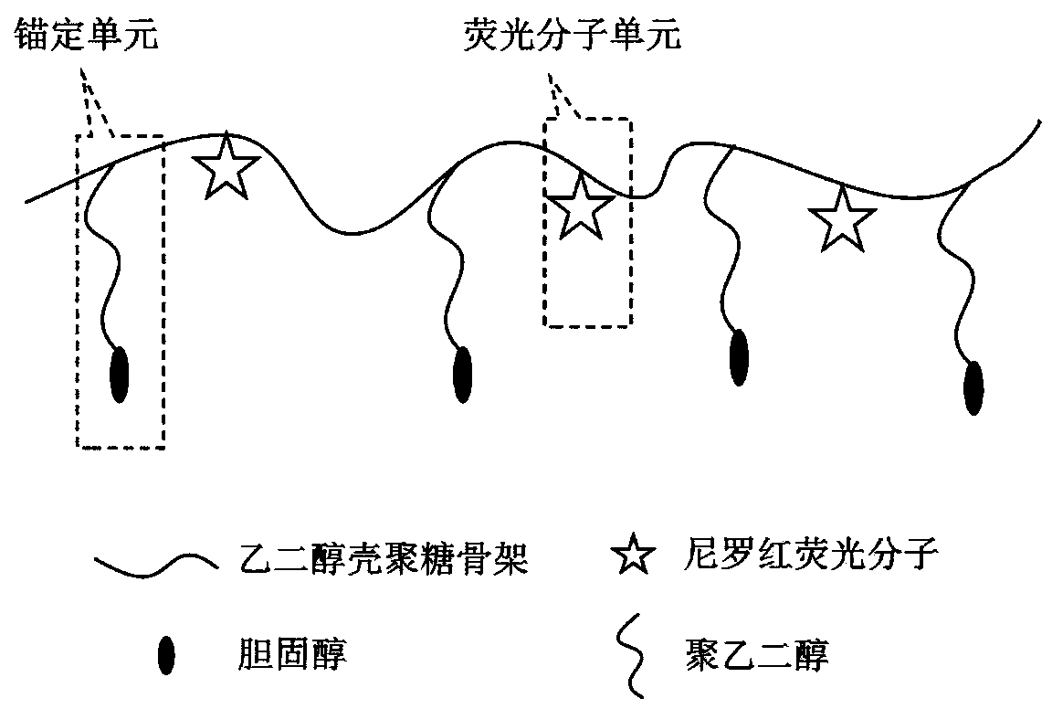
[0029] The design, synthesis and cell membrane fluorescence staining method of chitosan-30% cholesterol-2% Nile red, a multi-site anchoring, no-wash long-term cell membrane fluorescence imaging reagent, are as follows:

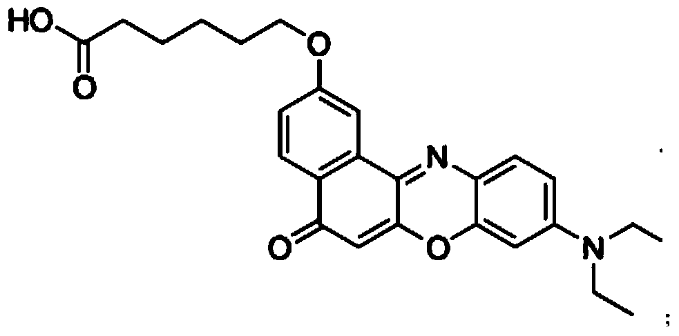
[0030] Reagent design: the reagent composition see figure 1 , The reagent uses glycol chitosan as the backbone, and the side chain contains 30% polyethylene glycol 2000-cholesterol (PEG2000-cholesterol) hydrophobic units and 2% Nile red dye fluorescent units. The ethylene glycol chitosan macromolecule has good biocompatibility and water solubility, and the molecular weight is above 10,000. The hydrophobic side chain of cholesterol is linked to the amino group of ethylene glycol chitosan in the form of NHS-PEG2000-cholesterol to increase water solubility. The Nile Red dye is carboxylated Nile Red, which is directly linked to ethylene glycol chitosan through reaction with amino groups. After the reagent is dissolved in water, it maintains a free stretching sta...

Embodiment 2

[0034] The implementation method of this embodiment is consistent with the method in embodiment 1, except that the Nile red fluorescent molecule is replaced by tetraphenylethylene (TPE) fluorescent molecule, and the molecular weight of the selected ethylene glycol chitosan is about 100,000. The fluorescent molecule is a kind of aggregation-induced emission (AIE), which can emit light through restriction of intra-molecular rotation (RIR). The fluorescent molecule has a large degree of freedom in aqueous solution, and the energy is easily dispersed, so the luminous efficiency is low; after being inserted into the hydrophobic cell membrane, because it is bound in the tight space of the hydrophobic phospholipid, the rotation of the molecule is limited, and the energy dispersion is hindered, resulting in Green fluorescence with high fluorescence quantum yield. Therefore, the luminescent condition of the fluorescent molecule is similar to that of Nile Red, and it can be used to make...

Embodiment 3

[0036] The implementation method of the present embodiment is similar to the method in Example 1, except that the high molecular weight of glycol chitosan selected is about 10000, and the percentage of synthetic anchor unit accounting for the number of repeating units of glycol chitosan is changed to 5%. , the percentage of fluorescent molecular units in the repeating units of ethylene glycol chitosan was changed to 1%, and the prepared imaging reagent consisted of chitosan-5% cholesterol-1% Nile red.


PUM


Abstract

The invention provides a washing-free long-time cell membrane fluorescence imaging reagent. The side chain of the reagent is an ethylene glycol chitosan macromolecule containing a hydrophobic unit and a low-efficiency fluorescence unit. Based on multi-site anchoring, the reagent has the advantages of good biocompatibility, simple synthesis, low cost and convenience in staining, does not need washing, cannot be easily endocytosed by cells, and can be used as a long-time cell membrane specificity tracing labelling probe.

Description

technical field [0001] The invention belongs to the technical field of biomedicine, and in particular relates to a long-time cell membrane fluorescence imaging reagent based on multi-site anchoring and no-cleaning, and also relates to a preparation method of the reagent. Background technique [0002] The cell membrane not only wraps individual cells structurally to form a stable internal environment, but also participates in material transport, energy transfer, signal transduction, vesicle transport, cell growth, differentiation, adhesion, transfer, stress, and endocytosis functionally , exocytosis, apoptosis, necrosis, autophagy and many other important biological processes, so more and more attention has been paid to the research on cell membranes. Fluorescent staining of cell membranes can be used to mark and track cell membranes, and has become a powerful method for studying the structure and function of cell membranes. [0003] Currently commercially available lipophil...

Claims


Application Information

Patent Timeline
Patent Type & Authority Applications(China)
IPC IPC(8): A61K49/00
Inventor 吴富根王宏银陈战
Owner SOUTHEAST UNIV
Why Eureka
Try Eureka