

Pepsinogen I detection method and kit thereof
A pepsinogen and detection method technology, applied in the field of pepsinogen I dual-wavelength fluorescence immunochromatography detection method and its detection kit, can solve the problems of long detection time, high detection results, complicated operation process, etc., and achieve The effect of reducing the influence of impurities
- Summary
- Abstract
- Description
- Claims
- Application Information
AI Technical Summary
Problems solved by technology
Method used
Image

Examples
Embodiment 1
[0035] The dual-wavelength fluorescent immunochromatographic detection method of pepsinogen I of the present invention comprises the following steps:
[0036] 1) Preparation of immunochromatographic test strips: Coat the pepsinogen Ⅰ monoclonal antibody and chicken IgY on the detection line (T line) and control line (C line) of the chromatography test paper, respectively, using a film spraying machine. After drying for 1-5 hours, cut with a strip cutter to obtain immunochromatographic test strips with a width of 3-5 mm;
[0037] It should be noted that: a) Traditional qualitative immunochromatography uses coated goat anti-mouse antibody as the control line. With the increase of pepsinogen Ⅰ or some anti-mouse antibody blocking agents in the serum, the signal on the control line As it decreases, its signal value cannot be used for calculation, and the accuracy of the signal of the test line has no reference basis. The present invention adopts chicken IgY antibody and goat anti...
Embodiment 2
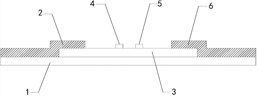
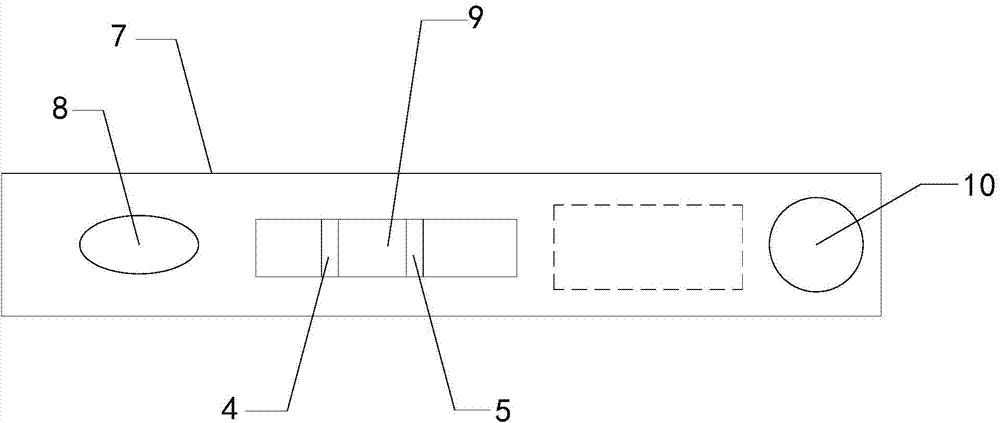
[0048] Such as Figures 1 to 2 As shown, the dual-wavelength fluorescent immunochromatographic detection kit of pepsinogen I of the present invention includes a kit body, a cryopreservation tube, and a diluent bottle, and the kit body includes a plastic liner 1 and is fixed on the plastic liner. The sample pad 2, the immunochromatography test strip 3 and the absorbent paper 6, the immunochromatography test strip is made of nitrocellulose membrane material, the sample pad and the absorbent paper are respectively lapped on both sides of the immunochromatography test strip, and the immunochromatography test strip is A detection line 4 and a control line 5 are set on the chromatography test strip, and the detection line and the control line are respectively coated with pepsinogen Ⅰ monoclonal antibody and chicken IgY; freeze-dried probes are stored in the cryopreservation tube, freeze-dried The probe is a freeze-dried probe prepared by reacting pepsinogen I monoclonal antibody and...
specific Embodiment
[0052]The present invention also provides a specific embodiment of dual-wavelength fluorescence immunochromatographic detection of pepsinogen I, which includes the following steps in sequence:
[0053] Step 1: Preparation of lyophilized probes
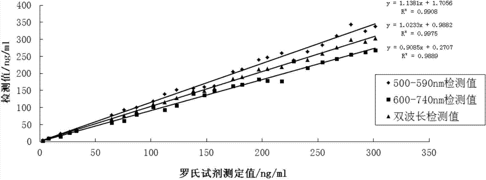
[0054] 1) Mix two fluorescent latex particle solutions with emission wavelengths of 550nm and 700nm respectively according to the volume ratio of 1:1. After mixing evenly, take 500 μl of mixed fluorescent latex particle solution (containing carboxyl groups) and use pH6.0 MES buffer After washing and centrifuging three times, the precipitate was diluted with pH6.0 MES buffer, and after adding 10mg EDC to mix well, the reaction was activated at room temperature for 30min. After centrifugation, the precipitate was washed three times with pH6.0 MES buffer, and then the precipitate was washed with pH6. Dilute with .0 MES buffer, add 125 μg pepsinogen Ⅰ monoclonal antibody, react at room temperature for 3 hours, add BSA to block, continue to...
PUM
| Property | Measurement | Unit |
|---|---|---|
| Width | aaaaa | aaaaa |
| Emission wavelength | aaaaa | aaaaa |
Abstract
Description
Claims
Application Information