Method for detecting chromosome microdeletion and micro-duplication of human embryo

A chromosomal micro-deletion and micro-duplication technology, applied in biochemical equipment and methods, microbial determination/inspection, etc., can solve the problems of low throughput, low resolution, high cost, etc., and achieve the effect of low cost and high sensitivity

Active Publication Date: 2015-07-01
BEIJING ZHONGYI KANGWEI MEDICAL INSTR
View PDF4 Cites 14 Cited by
  • Summary
  • Abstract
  • Description
  • Claims
  • Application Information

AI Technical Summary

Problems solved by technology
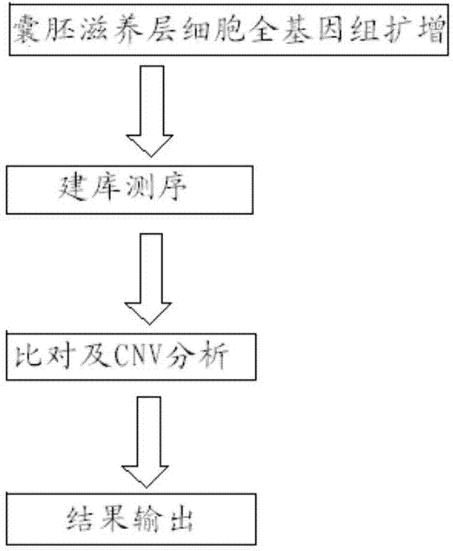
This method is simple and easy, but the disadvantage is that it can only detect known sites, and only a few sites can be detected at a time
[0012] Although, in 2014, Wells et al. reported the use of NGS technology for chromosome CNV detection and achieved full genome coverage, the resolution of this technology is still not high enough, and it can only detect chromosome aneuploidy and cannot detect chromosome microdeletions /microrepetition
The alg

Method used


Image

  • Method for detecting chromosome microdeletion and micro-duplication of human embryo
  • Method for detecting chromosome microdeletion and micro-duplication of human embryo
  • Method for detecting chromosome microdeletion and micro-duplication of human embryo

Examples


Embodiment 1

[0049] 1. Materials:

[0050] 1. Specimen: Blastocyst stage cells biopsied from a partner hospital. The source of the sample is the embryo samples from two cases of IVF. The parental karyotype examination is a balanced translocation carrier. Before this, an abnormal fetus was conceived and resulted in a miscarriage.

[0051] 2. Reagents: Qiagen's whole genome amplification kit, MDA product detection kit, MDA product purification kit, life library construction kit, Agencourt? AMPure? XP magnetic beads, life template preparation kit, life sequencing kit.

[0052] 3. Instruments: PCR instrument, agarose gel electrophoresis system, PGM sequencing platform of ion torrent.

[0053] 4. Consumables: 1.5ml, 0.2ml imported centrifuge tube, Ruining pipette and filter tip.

[0054] 2. Operation steps:

[0055] 1. Biopsy of trophectoderm cells

[0056] The two-step method is adopted. The first step is to punch the zona pellucida on D3 before biopsy. On D5 or D6, trophoblast cells are co...

Embodiment 2

[0153] 1. Materials:

[0154] 1. Specimen: Blastocyst stage cells biopsied by the cooperative hospital, the source of the sample is the embryo samples of three cases of IVF, the parents have suffered repeated miscarriage before.

[0155] 2. Reagents: Qiagen's whole genome amplification kit, MDA product detection kit, MDA product purification kit, life library construction kit, Agencourt? AMPure? XP magnetic beads, life template preparation kit, life sequencing kit.

[0156] 3. Instruments: PCR instrument, agarose gel electrophoresis system, PGM sequencing platform of ion torrent.

[0157] 4. Consumables: 1.5ml, 0.2ml imported centrifuge tube, Ruining pipette and filter tip.

[0158] 2. Operation steps:

[0159] 1. Biopsy of trophectoderm cells

[0160] The two-step method is adopted. The first step is to punch the zona pellucida on D3 before biopsy. On D5 or D6, trophoblast cells are collected from the 2 high-quality blastocysts formed under the microscope. Collect 5-10 tr...


PUM


Abstract

The invention relates to a method for detecting chromosome microdeletion and micro-duplication of a human embryo. The method comprises the following steps of performing whole genome amplification on cells cultured in vitro, interrupting DNA (deoxyribonucleic acid) molecules, and sequencing DNA fragments to obtain sequencing reads; comparing the sequencing reads with a reference sequence, and positioning the sequencing reads on the reference sequence; screening non-repeated areas of the reference sequence, and reserving the non-repeated areas; establishing a matrix of read number in windows through normal samples, analyzing the data of the normal samples, performing statistics on the read number of all the windows in the non-repeated areas, and establishing a probability matrix of the read number and chromosome enpeoids; calculating the copy number, i.e., the A/B/C state, of loci; selecting m continuous loci, i.e., the A state, as micro-duplication loci, and selecting m continuous loci, i.e., the C state, as microdeletion loci; contrasting the micro-duplication loci and the microdeletion loci with the existing CNV (copy number variation) and disease database, performing basic gene annotation and gene function analysis which relates to deletion parts, and annotating with a microdeletion syndrome disease type.

Description

technical field [0001] The invention relates to the field of molecular cell biology, in particular to a method for detecting microdeletions and microduplications of human embryonic chromosomes, which can detect the copy number variation of chromosomal DNA fragments of human embryonic blastocyst stage trophoblast cells. Background technique [0002] Chromosomal microdeletions / microduplications refer to deletions or duplications of 1.5kb-10Mb in length on a chromosome. Human chromosomal microdeletion / microduplication syndrome is a complex phenotypic disorder caused by deletions or duplications of tiny segments of human chromosomes, namely copy number variations of DNA segments, abbreviated as CNVs, which can lead to serious diseases and abnormalities such as congenital Sexual heart disease, growth retardation, limb deformity, etc. Common microdeletion syndromes include 22q11 microdeletion syndrome, catcalling syndrome, Angelman syndrome, AZF deletion, etc. [0003] The 200...

Claims


Application Information

Patent Timeline
IPC IPC(8): C12Q1/68
CPCC12Q1/6869C12Q1/6883
Inventor 冯涛杨凯邢丽贤
Owner BEIJING ZHONGYI KANGWEI MEDICAL INSTR
Why Eureka
Try Eureka