A biomimetic microfluidic chip for simulating tumor cells and their metastatic microenvironment in vivo

A technology of microfluidic chips and tumor cells, applied in the field of bionic microfluidic chips, can solve the problems of long experimental period, expensive animal models, inability to correctly reflect the physiological state of lung cancer cell metastasis, etc., and achieve good adaptability, good biological Compatibility, guaranteed gas exchange effect

Active Publication Date: 2017-06-30
THE SECOND HOSPITAL OF DALIAN MEDICAL UNIV
View PDF6 Cites 0 Cited by
  • Summary
  • Abstract
  • Description
  • Claims
  • Application Information

AI Technical Summary

Problems solved by technology

However, currently available animal models for evaluating normal physiological and disease processes are expensive, subject to lengthy experimental cycles, and present various ethical controversies
More importantly, the above-mentioned animal models cannot well control the location and tropism of metastatic lung cancer cells, resulting in the inability to correctly reflect the physiological state of lung cancer cell metastasis in humans.

Method used


Image

  • A biomimetic microfluidic chip for simulating tumor cells and their metastatic microenvironment in vivo
  • A biomimetic microfluidic chip for simulating tumor cells and their metastatic microenvironment in vivo
  • A biomimetic microfluidic chip for simulating tumor cells and their metastatic microenvironment in vivo

Examples


Embodiment 1

[0058] Example 1 A bionic microfluidic chip for simulating the microenvironment of lung cancer metastasis in vivo

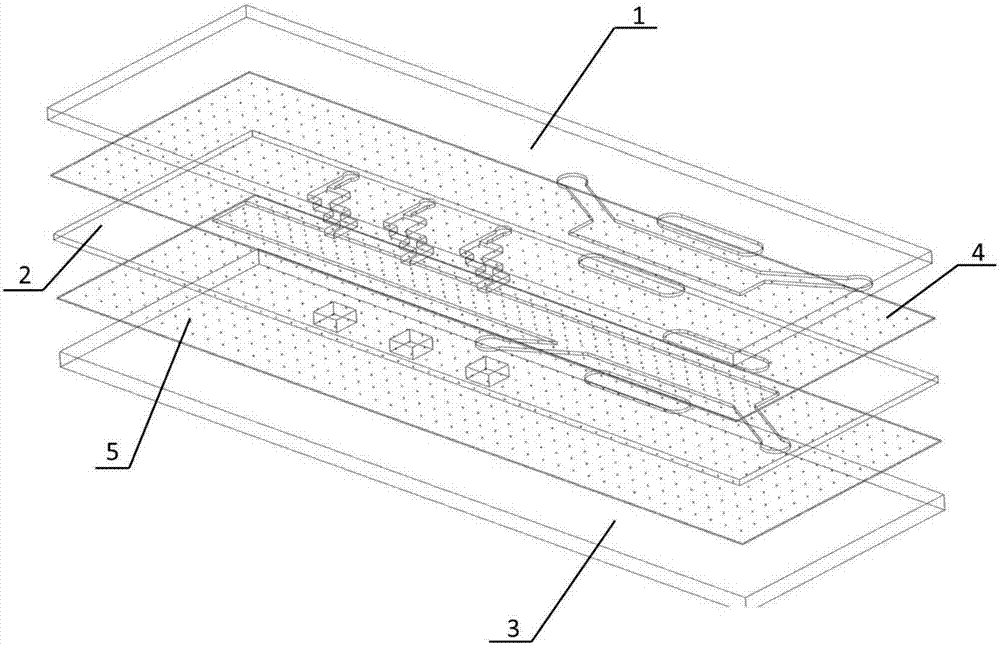
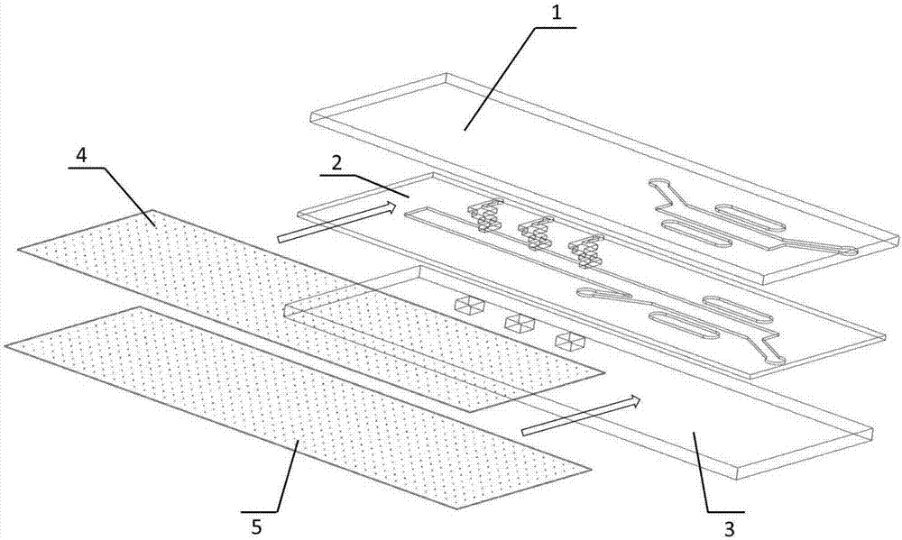
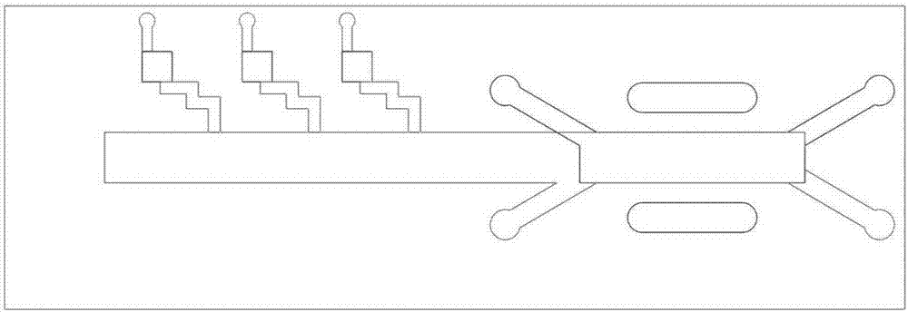
[0059] The microfluidic chip of the present invention is an airtight whole formed by interlacing and irreversible sealing of three layers of PDMS substrates and two layers of porous PDMS membranes; the first layer of PDMS substrate 1 is provided with an air channel 11 for air circulation , the first layer of liquid inlet 12 and the first layer of liquid outlet 13 for liquid to enter and exit, respectively located in the upper half of the first vacuum channel 14 and the second vacuum channel 15 on both sides of the air channel 11; the second layer of PDMS substrate 2 A liquid passage 21 for liquid circulation, a second-layer liquid inlet 22 and a second-layer liquid outlet 23 for liquid to flow in and out are arranged on the top, and are respectively located under the first vacuum passage 14 and the second-layer liquid 15 on both sides of the liquid passage 21. In...

Embodiment 3

[0073] Example 3 Construction of a bionic model simulating the microenvironment of lung cancer cell metastasis in vivo

[0074] 1. 2D culture of lung cancer cells in a microfluidic chip

[0075] (1) The microfluidic chip is sterilized by ultraviolet irradiation, and the porous PDMS membrane is coated with BME. The specific coating process is: pretreatment of the chip, so that cells can better adhere to the porous membrane surface of the chip. Dilute BME (Cultrexbasement membrane extract, R&D Systems, McKinley Place, MN, USA) at a ratio of 1:10, mix well, inject into the sample inlet of the microfluidic chip with a micro-sampler, and wait overnight in the incubator for the gel to solidify.

[0076] (2) The microfluidic chip is turned over, that is, the third PDMS substrate faces upward. Collect the suspended mononuclear cells into a centrifuge tube, centrifuge at 1000 rpm for 5 minutes, discard the supernatant, and add fresh medium to prepare a cell suspension. The mononuclea...

Embodiment 4

[0087] Example 4 Detection of the effectiveness of the bionic model for simulating the metastatic microenvironment of lung cancer cells in vivo

[0088] Evaluate the effectiveness of the bionic model of the simulation body lung cancer cell transfer microenvironment constructed in Example 3, and complete it through the following experiments:

[0089] 1. Detection of cell viability

[0090] Detection method: absorb the culture medium in the channel in the chip system, inject PBS into the chip channel, and wash the cells of different treatment groups twice; then pump H33342 (1:100) into the staining for 15 minutes, and wash twice with PBS solution; Pump in PI staining (1:200) for 5 minutes, wash with PBS solution twice; under a microscope, observe the fluorescence intensity under the excitation of the corresponding excitation light and record it by taking pictures.

[0091] 2. Detection of co-localization of lung cancer cells and bronchial epithelial cells

[0092] The cell tra...


PUM

PropertyMeasurementUnit
lengthaaaaaaaaaa
lengthaaaaaaaaaa
pore sizeaaaaaaaaaa

Abstract

The invention discloses a bionic micro-fluidic chip for simulating in vivo tumor cells and their metastasis microenvironment. The micro-fluidic chip is formed by interlacing and irreversible sealing of three layers of PDMS substrates and two layers of porous PDMS membranes. The first PDMS substrate layer is provided with a channel for air circulation, an inlet and an outlet for entry and exit of a liquid and an upper half part of a vacuum channel. The third PDMS substrate layer is provided with a cell culture room for cell culture. The channel of the first PDMS substrate layer is positioned up the channel of the second PDMS substrate layer. The cell culture room on the third PDMS substrate layer is positioned down the channel of the second PDMS substrate layer. The micro-fluidic chip provided by the invention can be used for in vitro detection of the tumor cell metastasis process and provides a basis for making clinical tumor treatment plans.

Description

technical field [0001] The invention belongs to the field of clinical application, and relates to a bionic microfluidic chip for simulating the microenvironment of tumors and their metastases in the body. The microfluidic chip of the present invention can dynamically monitor the metastasis process of tumor cells, define the migration mode of tumor cells, and provide guidance for preventing and treating tumor metastasis. Background technique [0002] Cancer cell metastasis is the main cause of death from all cancers, including lung cancer, and is one of the biggest challenges for basic researchers and clinical scholars. Cancer cell metastasis is a complex physiological process that usually includes primary site tumor lesion proliferation, tumor cell detachment, tumor cell migration, extravasation, and formation of metastatic tumor cell clusters at second organs. Tumor cells with a metastatic phenotype exhibit the following properties: enhanced cell mobility, increased abilit...

Claims


Application Information

Patent Timeline
Patent Type & Authority Patents(China)
IPC IPC(8): C12M3/00C12Q1/02G01N33/68
CPCB01L3/5027B01L3/502707C12M23/16C12M35/08G01N33/5005
Inventor 王琪高占成许志赟郭哲郝华龙徐一彤
Owner THE SECOND HOSPITAL OF DALIAN MEDICAL UNIV
Why Eureka
Try Eureka