Circulating tumor cell detection kit, preparing method thereof and application thereof

A technology for detecting kits and tumor cells, applied in the field of circulating tumor cell detection kits and their preparation, can solve the problems of low capture efficiency and short shelf life of the detection kits, and achieve long storage time, easy storage, and increased collision probability. Effect

Inactive Publication Date: 2015-12-02
SHANGHAI JIAO TONG UNIV
View PDF5 Cites 39 Cited by
  • Summary
  • Abstract
  • Description
  • Claims
  • Application Information

AI Technical Summary

Problems solved by technology

[0006] The invention provides a circulating tumor cell detection kit and its preparation method and application,

Method used


Image

  • Circulating tumor cell detection kit, preparing method thereof and application thereof
  • Circulating tumor cell detection kit, preparing method thereof and application thereof
  • Circulating tumor cell detection kit, preparing method thereof and application thereof

Examples


Embodiment 1

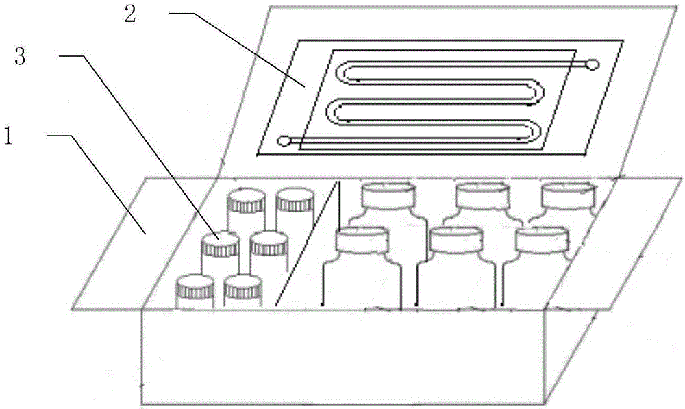
[0047] Such as figure 1 As shown, the circulating tumor cell detection kit includes a box body 1, a microfluidic chip 2 arranged in the box body 1, and a reagent bottle set 3; the reagent bottle set includes a cell recognition probe, red blood cell lysate , Washing Solution, Blocking Solution, Fixative Solution, Permeabilization Solution, and Fixed Cell Stain.

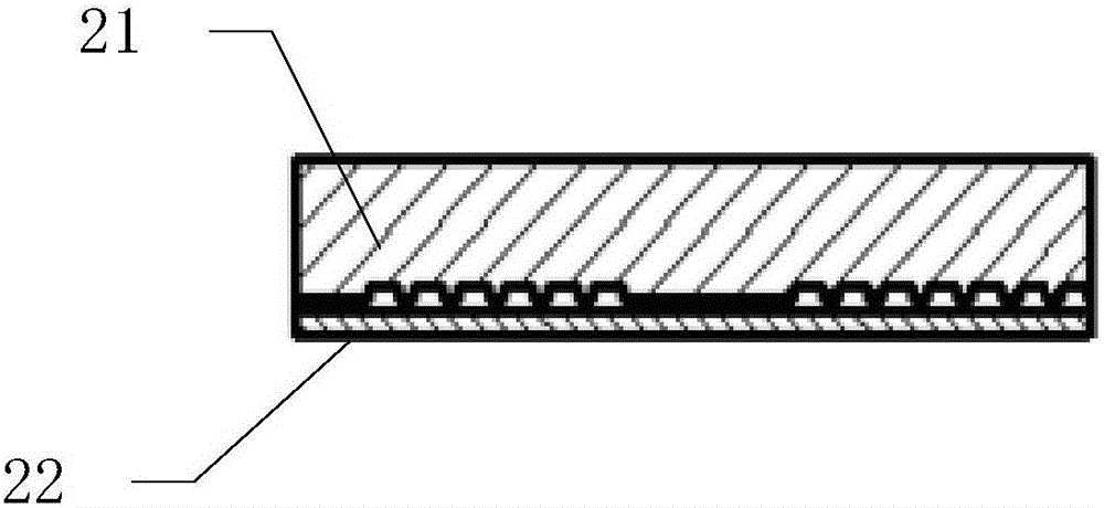
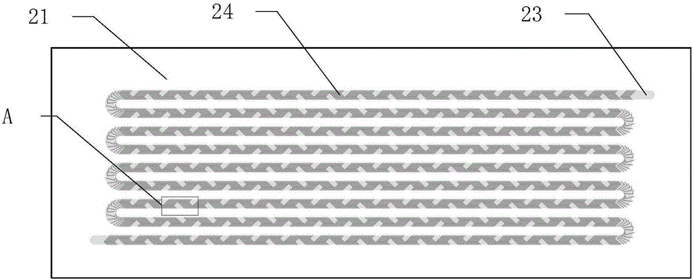
[0048] Such as figure 2 As shown, the microfluidic chip 2 is composed of a PDMS substrate 21 and a slide glass 22, as image 3 As shown, microchannels 23 are distributed on the surface of the PDMS substrate 21, the microchannels 23 are single channels, and are arranged in 9 rows in a continuous S-shape, and the bottom surface of the microchannels 23 has a periodic interlaced mixing structure 24, Such as Figure 4 As shown, the interlaced mixing structure 24 is a herringbone structure.

[0049] Such as Figure 5 As shown, after the PDMS substrate 21 is bonded to the slide glass 22, the microfluid channel 25 is for...

Embodiment 2

[0056] The difference from Example 1 is that the reagent bottle set includes a cell recognition probe reagent bottle 31 , a red blood cell lysate reagent bottle 32 , a cleaning solution reagent bottle 33 , a blocking solution reagent bottle 34 and an unfixed cell staining reagent bottle.

[0057] In addition, the slide 22 is a polylysine-modified slide from ThermoScientific, and the capture probe 26 also modifies the surface of the slide 22 in addition to the surface of the microchannel 23 .

[0058] The non-fixable cell stain includes Cy3-labeled first single-stranded polynucleotide and APC-labeled anti-CD45. Sequentially select the filter slides of CY5 and TRITC to observe under a fluorescent microscope, the first single-stranded polynucleotide labeled with Cy3 is red, and the APC-labeled anti-CD45 is green.

Embodiment 3

[0059] The preparation method of the microfluidic chip described in embodiment 3

[0060] The preparation method of the microfluidic chip 2 described in Example 1 and Example 2 is as follows: After the PDMS substrate 21 is treated with oxygen in a plasma cleaner for 15 seconds, it is soaked in a 4% 3-aminopropyltriethoxysilane solution. Soak for 1 hour to generate amino groups on the surface, and then wash with ultrapure water to remove excess reagents.

[0061] After drying with nitrogen gas, the amino-modified PDMS substrate 21 was attached to a clean glass slide 22, and placed in an oven at 80° C. for 2 hours, and after returning to room temperature, one-step loading and capturing anti-probe 26 was carried out. The specific method is: mix 500 μL of amino-modified first single-stranded polynucleotide with DMSO at a volume ratio of 3:2, and then mix the mixed solution with BS3 (disuccinimide suberate sodium salt) aqueous solution at a volume ratio of 1:1. After mixing, pass ...


PUM

PropertyMeasurementUnit
Bump heightaaaaaaaaaa
Heightaaaaaaaaaa
Widthaaaaaaaaaa

Abstract

The invention particularly relates to a circulating tumor cell detection kit, a preparing method thereof and an application thereof. The circulating tumor cell detection kit comprises a kit body, detection reagents and a micro-fluidic chip, wherein the detection reagents and the micro-fluidic chip are arranged in the kit body. The micro-fluidic chip is formed by a PDMS substrate and a glass slide in an attached mode. A micro-channel is distributed on the surface of the PDMS substrate, and a fishbone-shaped alternate type mixing structure is arranged on the bottom face of the micro-channel. After the PDMS substrate and the glass slide are bonded, a microfluid channel is formed on the surface of the micro-channel and the surface of the glass slide, and capturing probes are loaded on the surface of the micro-channel. The invention further provides the preparing method of the micro-fluidic chip and a method for detecting circulating tumor cells through the kit. Compared with the prior art, the circulating tumor cell detection kit and the methods have the advantages that the capture probes are loaded on all the surfaces of the micro-channel, and therefore the capturing efficiency is improved; as the micro-fluidic chip is modified through single strand nucleotide, the storage time of the chip is prolonged; in addition, a cell-fixing-free straining method is provided, and therefore convenience is provided for further cell gene sequencing.

Description

technical field [0001] The invention belongs to the field of rare cell detection, and in particular relates to a circulating tumor cell detection kit and its preparation method and application. Background technique [0002] Circulating tumor cells in human peripheral blood are a kind of rare cells that exist in the peripheral blood of cancer patients with a small number but have important clinical significance. They are tumor cells that spread into the peripheral blood circulation from tumor lesions and can develop into metastatic lesions. Circulating tumor cells in the blood suggest the existence of tumors and possible metastasis in the body, and more than 90% of cancer deaths in clinical practice are caused by metastasis. Therefore, capturing and detecting circulating tumor cells from blood has attracted more and more attention. [0003] The amount of circulating tumor cells in peripheral blood is very small, and every 10ml of blood may contain only a few to dozens of ci...

Claims


Application Information

Patent Timeline
IPC IPC(8): G01N15/10G01N33/574
Inventor 施奇惠邓宇亮汪民娇王智华
Owner SHANGHAI JIAO TONG UNIV
Why Eureka
Try Eureka