
Preparation method and application of multi-epitope fusion diagnostic antigen protein of Echinococcus granulosus

A technology for Echinococcus granulosus disease and fusion antigen, which is applied in the field of Echinococcus granulosus multi-epitope fusion diagnosis antigen protein and its preparation, and can solve the problems of poor specificity and weak reactogenicity.

Inactive Publication Date: 2019-06-21
SHIHEZI UNIVERSITY
View PDF3 Cites 0 Cited by
  • Summary
  • Abstract
  • Description
  • Claims
  • Application Information

AI Technical Summary

Problems solved by technology

[0006] Studies have shown that although Eg cyst fluid diagnostic antigen has high sensitivity, its specificity is poor, and it has a high degree of cross-reaction with other taeniasis sera; although Eg recombinant single protein antigen has high specificity, the reactant Weaker

Method used


Image

  • Preparation method and application of multi-epitope fusion diagnostic antigen protein of Echinococcus granulosus
  • Preparation method and application of multi-epitope fusion diagnostic antigen protein of Echinococcus granulosus
  • Preparation method and application of multi-epitope fusion diagnostic antigen protein of Echinococcus granulosus

Examples


Embodiment 1

[0038] In this example, sheep from a pasture in Xinjiang were used as specimens, and Eg scoliosis of sheep was aseptically cultured and collected. Other raw materials were obtained by purchasing kits and the like.

[0039] 1. Cloning of Eg important antigen gene

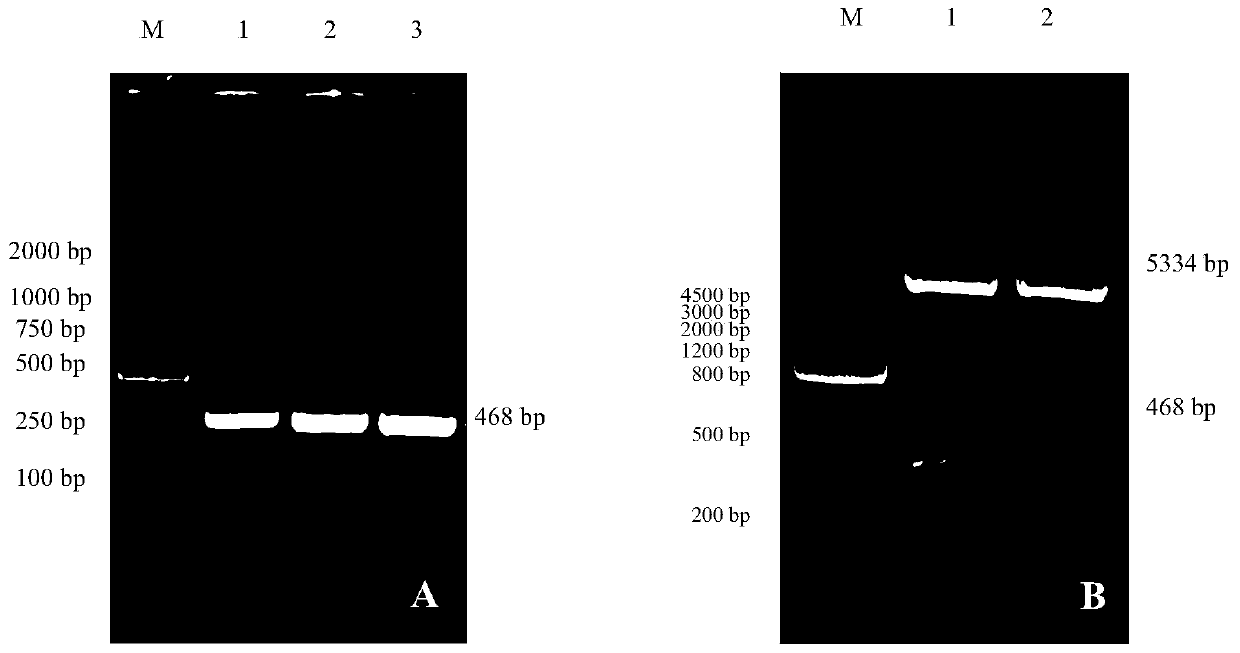
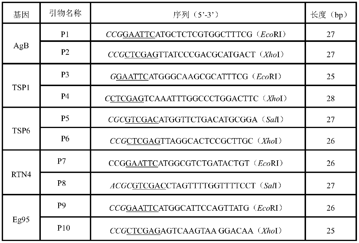
[0040] Specific primers were designed according to the cDNA sequences of Eg AgB (AY871041.1), TSP1 (EG_11043), TSP6 (EG_00715) and RTN4 (EG_04657) genes, and the total RNA template of sheep Eg scole was used for RT-PCR amplification. The PCR product was cloned into the pMD19-T vector, and after sequencing, bioinformatics analysis was performed on the above gene sequence.

[0041] 2. Prediction analysis of Eg antigen protein T cell epitope

[0042] Using biological online software to predict and analyze the epitopes of the amino acid sequences encoded by the important genes of Eg (AgB, TSP1, TSP6, RTN4 and Eg95), and combine four different prediction methods to find the sequences with common dominant linear epitopes ...

Embodiment 2

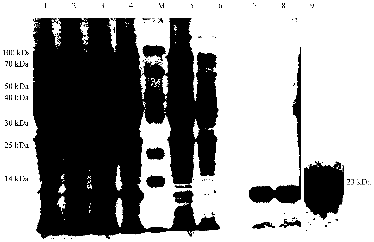
[0054] 1. Establishment of indirect ELISA method and condition optimization based on Eg-meAg1 antigen protein

[0055] (1) Determination of the best working conditions: Dilute the purified protein with 0.05M pH 9.6 carbonate buffer, and the antigens are serially diluted to 64, 32, 16, 8, 4 and 2ug / mL, including 96-well ELISA plates were coated with 100 μL per well. Coating conditions: After the ELISA plate is coated, overnight at 4°C, take it out, wash with PBST 3 times, 3min each time; use 1% gelatin, 5% skimmed milk powder and 5% BSA as the blocking solution, 200μL / well, 37°C respectively Blocked for 0.5h, 1h, 1.5h and 2h, washed 3 times; Eg positive serum was diluted 1:50, 1:100 and 1:200 respectively, 100μL / well, serum action time was set to 1h, the same sample was used as 3 replicate wells, at the same time as a blank control well, washed 3 times; the enzyme-labeled antibody was first diluted 1:2 500, 1:5 000 and 1:10 000, 100 μL / well, and reacted at 37°C for 0.5h and 1h...


PUM


Abstract

The invention relates to a multi-epitope fusion antigen protein for echinococcosis granulosis cyst diagnosis and a preparation method and application of the multi-epitope fusion antigen protein. The method comprises the steps that protein genes with echinococcosis granulosis cyst immunogenicity are screened out, dominant linear epitopes of the antigen gene coding protein is analyzed by means of the bioinformatics means, the epitopes are connected in series according to a certain sequence, the multi-epitope fusion antigen gene Eg-meAgl is constructed and expressed in escherichia coli, and the recombinant antigen protein Eg-meAgl with the diagnostic value and high immuneoreactivity is obtained. The prepared echinococcosis granulosis cyst recombinant multi-epitope fusion antigen Eg-meAgl has the high specificity and sensitivity, and is the valuable Eg infection diagnosis antigen, and a novel method for antigen protein preparation is provided for echinococcosis granulosis cyst (echinococcosis) diagnosis.

Description

technical field [0001] The invention relates to the technical field of biological detection, in particular to an antigenic protein for the diagnosis of Eg (Echinococcus granulosus) infection, in particular to a multi-epitope fusion diagnostic antigenic protein for Echinococcus granulosus and its preparation method and application . Background technique [0002] Echinococcus granulosus is distributed worldwide and is widely prevalent in countries such as the Middle East, Central Asia, Latin America, Australia, and southern Europe. It is also prevalent in 21 provinces (municipalities) and autonomous regions in my country, especially in Xinjiang, Gansu, Ningxia, Pastoral or semi-pastoral areas in Qinghai, Tibet, Sichuan and Inner Mongolia provinces are most severely affected. [0003] The economic loss of my country's livestock products due to the infection of echinococcus in animals has exceeded 3 billion yuan, seriously affecting the production and development of animal husba...

Claims


Application Information

Patent Timeline
IPC IPC(8): C07K19/00C12N15/62C12N15/70G01N33/68
CPCC07K14/4355C07K2319/00C12N15/70C12N2800/101C12N2800/22G01N33/68G01N2333/43539
Inventor 孟庆玲刘田莉乔军乔梦凡陈英贡莎莎田路路李重阳孟丹于伟伟陈创夫
Owner SHIHEZI UNIVERSITY
Learn More