Gas-liquid interface three-dimensional culture method for skin and hair follicle regeneration
A gas-liquid interface, three-dimensional culture technology, applied in biochemical equipment and methods, 3D culture, tissue culture and other directions, can solve the problems of wasting time and material resources, easily affected by the external environment, instability and other problems, saving time and material resources , Reasonable design, simple operation effect
- Summary
- Abstract
- Description
- Claims
- Application Information
AI Technical Summary
Problems solved by technology
Method used
Image
Examples
Embodiment 1
[0061] (1) Fabrication of gas-liquid interface support:
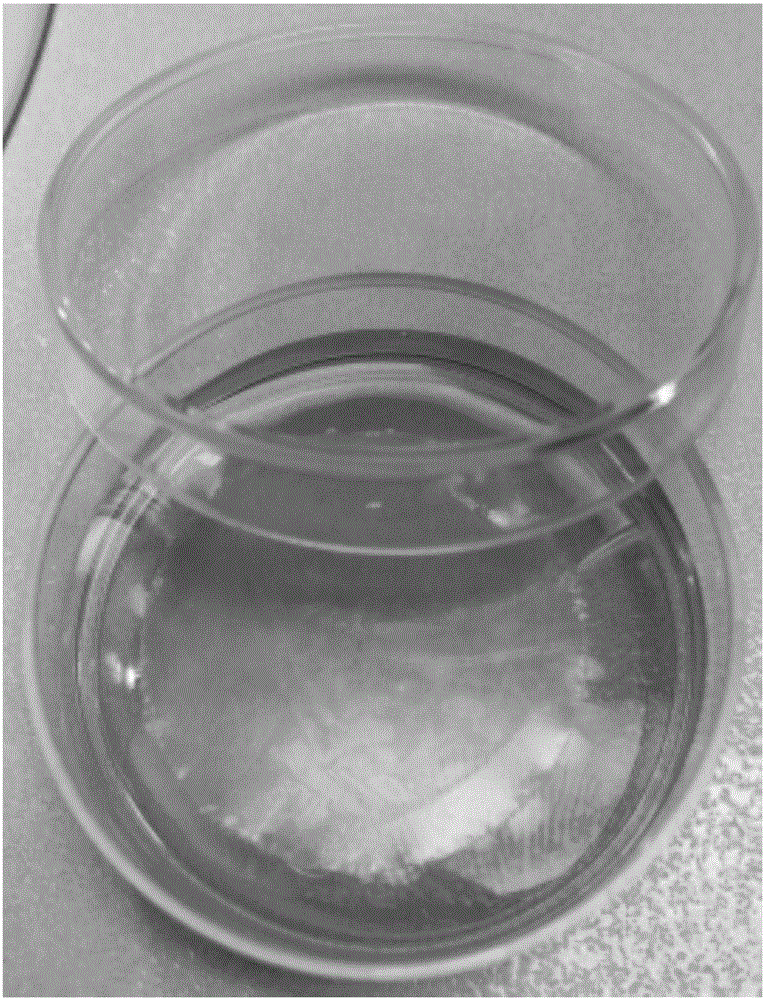
[0062] Nuclepore polycarbonate membrane, made of high-quality polycarbonate film, each Nucle-pore polycarbonate membrane is round, with different specifications and different pore diameters, use 40 μL rat tail type I collagen mixed with the same amount of PBS, sterile Spread evenly on the inner wall of a disposable 90mm-diameter petri dish under certain conditions, pick up the Nuclepore polycarbonate membrane with sterile ophthalmic forceps, spread it on a petri dish coated with rat tail collagen, seal the petri dish with a parafilm, and irradiate with ultraviolet light After 30 minutes, the rat tail type I collagen and the Nuclepore polycarbonate membrane were naturally dried, and then placed in a 4°C refrigerator for later use.
[0063] (2) Preparation of culture medium:
[0064] In the experiment, low-sugar MEM was used as the basic culture medium, and fetal calf serum (FCS) was added to make a 5% FCS+MEM culture me...
Embodiment 2
[0070] Get pregnant mice whose gestational age is between 14-17d (E14-E17), kill them by neck dislocation, soak in 75% ethanol for 10 min, wash 3 times in sterile PBS, and put The uterus was taken out and washed 3 times in sterile PBS to remove residual blood and mucus on the surface. After the uterus was cut open, the embryos with fetal membranes were removed and washed 3 times in sterile PBS. Tear the fetal membrane with forceps and take out the fetal mouse. Rinse the fetal mouse in sterile PBS and cut it with ophthalmic scissors along the two sides of the back, carefully peel off the back skin of the fetal mouse with tweezers, and cut the embryonic skin into a regular quadrilateral with a micro-ophthalmological scalpel. And use a puncher to create an incision in the center of the quadrilateral skin, measure and record the diameter of the incision.
[0071] Add 2mL of the prepared culture solution into a 30mm petri dish, float the treated Nucle-pore polycarbonate membrane ...
Embodiment 3
[0073] On the Nuclepore polycarbonate membrane, add 2 μL of stem cell proliferation marker EdU dropwise, use sterile ophthalmic forceps to transfer the dissected outer root sheath of the hair follicle to the floating Nuclepore polycarbonate membrane, and remove the hair follicle outer root sheath and stem cell proliferation marker Fully exposed to EdU, placed at 37°C, 5% CO 2 Cultured in the incubator for 12 h, immunofluorescence staining was used to observe the proliferation state of hair follicle stem cells ( Figure 8 ).
PUM
 Abstract
Description
Claims
Application Information
 - R&D
- Intellectual Property
- Life Sciences
- Materials
- Tech Scout
- Unparalleled Data Quality
- Higher Quality Content
- 60% Fewer Hallucinations
Browse by: Latest US Patents, China's latest patents, Technical Efficacy Thesaurus, Application Domain, Technology Topic, Popular Technical Reports.
© 2025 PatSnap. All rights reserved.Legal|Privacy policy|Modern Slavery Act Transparency Statement|Sitemap|About US| Contact US: help@patsnap.com


