Coxsackievirus A6 type infected animal model as well as preparation method and application thereof

A technology for coxsackie virus and infecting animals, applied in the biological field, can solve the problems of lack of stable animal infection models, constraints on the development of anti-CVA6 drugs and vaccines, and unclear pathogenesis of hand-foot-mouth disease

Inactive Publication Date: 2017-06-20
TAISHAN MEDICAL UNIV
View PDF0 Cites 13 Cited by
  • Summary
  • Abstract
  • Description
  • Claims
  • Application Information

AI Technical Summary

Problems solved by technology

At present, the pathogenesis of hand, foot and mouth disease is still unclear, especially the pathogenic mechanism of CVA6 has not been reported, and the screening of anti-CV

Method used


Image

  • Coxsackievirus A6 type infected animal model as well as preparation method and application thereof
  • Coxsackievirus A6 type infected animal model as well as preparation method and application thereof
  • Coxsackievirus A6 type infected animal model as well as preparation method and application thereof

Examples


Embodiment 1

[0043] Example 1, Isolation and Identification of Coxsackievirus Type A6 (CVA6) Strain WF057R

[0044] In 2015, the inventor isolated a Coxsackievirus type A6 (Coxsackievirus) strain from the feces of a hand-foot-mouth child in Shandong Province, and named the strain WF057R. Coxsackievirus type A6 (Coxsackievirus) strain WF057R has been preserved in the General Microbiology Center of China Committee for the Collection of Microbial Cultures (CGMCC for short) on December 26, 2016. The address is: Courtyard 3, No. 1 Beichen West Road, Chaoyang District, Beijing No.), the deposit number is CGMCC No.13393.

Embodiment 2

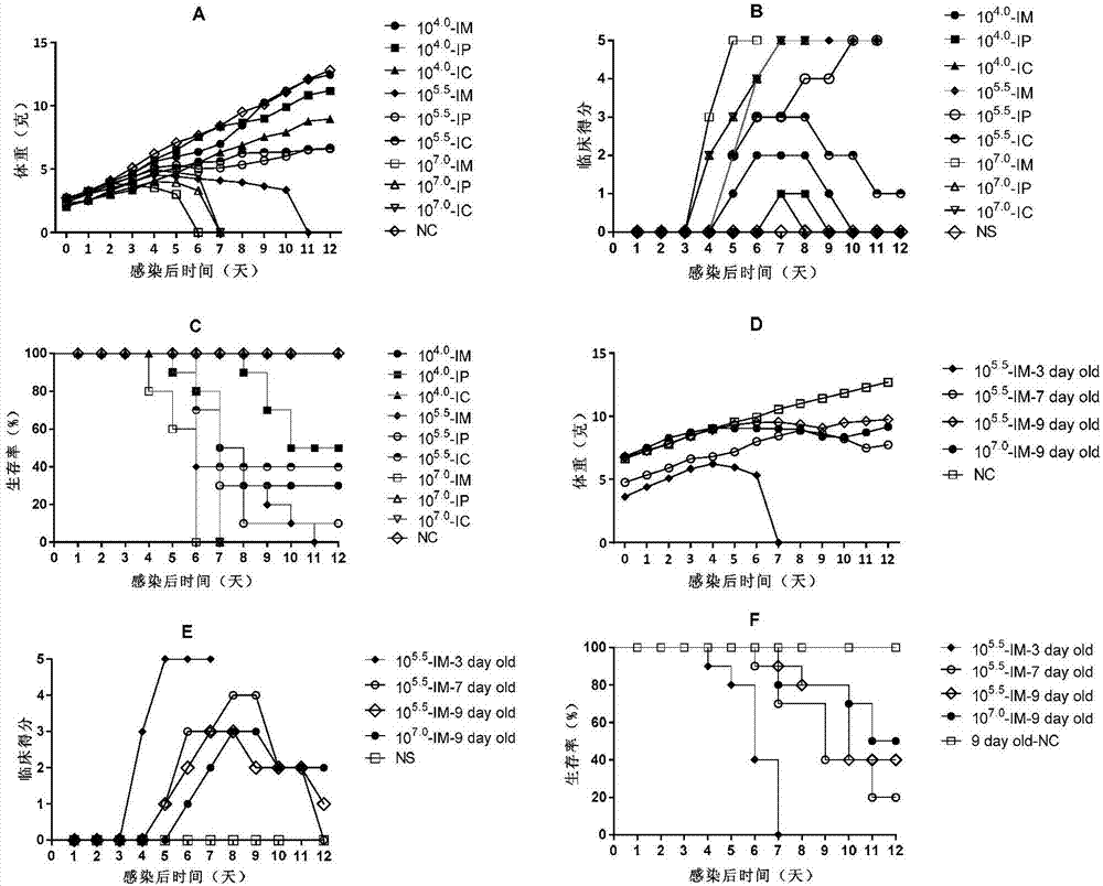
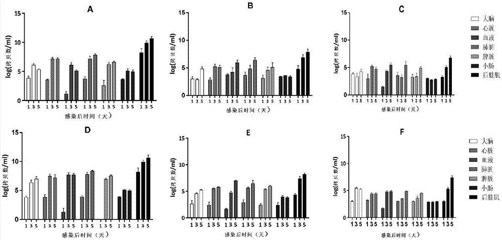
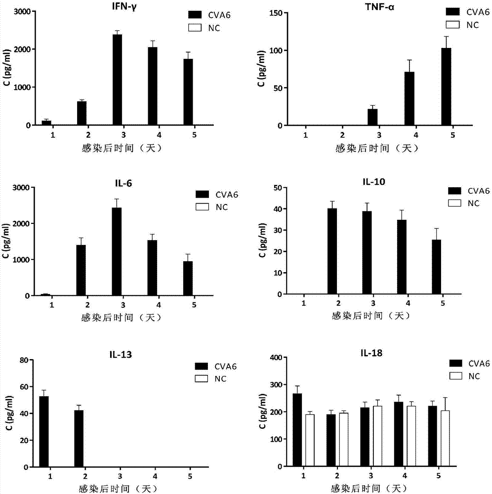
[0045] Example 2, Utilizing Coxsackie A6 Virus Strain WF057R to Prepare CVA6 Infected Animal Model

[0046] 1. Cultivation of WF057R

[0047] RD cells were inoculated in medium 1 (medium 1 is the liquid obtained by adding fetal bovine serum, penicillin and streptomycin to MEM maintenance solution, wherein the mass percentage concentration of fetal bovine serum is 10%, and the mass percentage of penicillin Concentration is 1%, the mass percent concentration of streptomycin is 1%), in temperature is 37 ℃, 5% concentration CO 2 cultured in a cell incubator to obtain RD cell culture medium. The Coxsackie A6 virus strain WF057R of Example 1 was inoculated on the RD cells in the RD cell culture medium, and when the cells had a cytopathic effect (Cytopathic effect, CPE) area exceeding 80%, the cell culture medium was collected to obtain CVA6 strain WF057R virus liquid, quantify virus by limiting dilution method in 96-well plate, and measure virus TCID 50 Afterwards, it was aliquot...

Embodiment 3

[0079] Example 3. The antiserum produced by the active CVA6 vaccine using inactivated WF057R can protect mice and treat diseases caused by CVA6

[0080] 1. Preparation of inactivated CVA6 vaccine

[0081] The diluted CVA6 strain WF057R virus liquid (10 5 TCID 50 / ml) was diluted with formalin at a ratio of 1:4000 (V / V) to obtain a virus dilution, and the virus dilution was incubated at 37°C for 72 hours to obtain an inactivated virus solution; the inactivated virus mixed with complete Freund's adjuvant in equal volumes to make a complete emulsion, which is the CVA6 vaccine, and the content of inactivated virus in the CVA6 vaccine is 5×10 4 TCID 50 / ml. The infectious titer of CVA6 vaccine was detected by micro titration method to observe its inactivation effect.

[0082] Virus micro titration method: 1×10 per well 4 Cell / 100 μ l culture fluid (the cell in this culture fluid is RD cell, and substratum is the substratum 1 of embodiment 2) RD cell is inoculated on 96 wells ...


PUM


Abstract

The invention discloses a coxsackievirus A6 type infected animal model as well as a preparation method and an application thereof. The preparation method of the coxsackievirus A6 type infected animal model provided by the invention comprises a step of infecting an animal with an inactivated coxsackievirus A6 type strain, so that the coxsackievirus A6 type infected animal model is obtained, wherein the coxsackievirus A6 type strain is preserved in China General Microbiological Culture Collection Center with preservation number of CGMCC No.13393. The coxsackievirus A6 type infected animal model, which is stable and is good in repeatability, is constructed on the basis of the CVA6 strain WF057R; and with the application of the model, a research tool is provided for next medicine antiviral therapy and assessment of an immune protective effect of an inactivated viral vaccine.

Description

technical field [0001] The invention relates to a Coxsackievirus A6 infection animal model and a preparation method and application thereof in the field of biotechnology. Background technique [0002] Coxsackievirus (CV) is a single-stranded positive-sense RNA virus belonging to the family Picornaviridae and the genus Enterovirus. Coxsackievirus particles are icosahedral stereosymmetric, spherical in shape, about 23-33nm in diameter, nucleocapsid exposed, without envelope and protrusions; Coxsackievirus genome contains only one open reading frame (ORF), encoding a relatively long polyprotein precursors. The ORF is sequentially divided into three regions, P1, P2 and P3, wherein the P1 region encodes the structural proteins VP1-VP4 of the virus, which constitute the viral capsid. The P2 and P3 regions encode nonstructural proteins. [0003] Coxsackie virus is the main pathogen that causes hand, foot and mouth disease (HFMD) in children, especially infants under 3 years old,...

Claims


Application Information

Patent Timeline
IPC IPC(8): C12N7/00A01K67/027C12R1/93
CPCA01K67/027A01K2207/20A01K2227/105A01K2267/0337C12N7/00C12N2770/32321
Inventor 张振杰史卫峰董兆鹏
Owner TAISHAN MEDICAL UNIV
Why Eureka
Try Eureka