Method of detecting adenosine with fluorescent sensor on the basis of aptamer

An aptamer sensor and fluorescence sensor technology, applied in the field of analysis and detection, can solve the problems affecting the sensitivity and selectivity of the sensor, and achieve the effects of facilitating in vitro screening and preparation, reducing background fluorescence and high specificity

Inactive Publication Date: 2017-07-14
NANJING MEDICAL UNIV
View PDF5 Cites 29 Cited by
  • Summary
  • Abstract
  • Description
  • Claims
  • Application Information

AI Technical Summary

Problems solved by technology

As a new type of fluorescent nanomaterial, carbon dots have been widely used in various fields. However, most of the current fluorescent sensors are analyzed

Method used


Image

  • Method of detecting adenosine with fluorescent sensor on the basis of aptamer
  • Method of detecting adenosine with fluorescent sensor on the basis of aptamer
  • Method of detecting adenosine with fluorescent sensor on the basis of aptamer

Examples


Embodiment 1

[0034] a) Construction of fluorescent aptamer sensor: Mix 45 μLaptamer-AuNPs and 30 μL ssDNA-CDs evenly, add 105 μL 10 mmol / L pH7.4 phosphate buffer, and incubate at 37°C for 30 min;

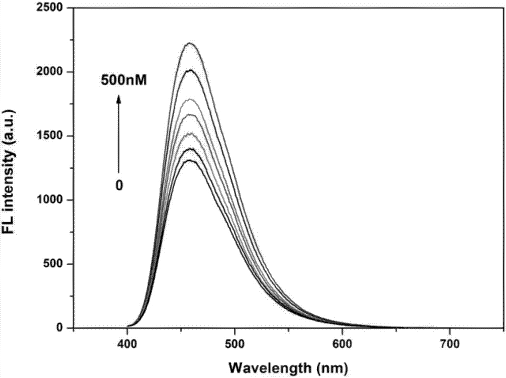
[0035] b) Detection of adenosine by fluorescent sensing: Add 20 μL of OnM, 0.1 μM, 0.25 μM, 1.5 μM, 2.5 μM, 3.5 μM, 5 μM adenosine solutions to the sensor constructed in step a), incubate at room temperature for 30 min, and measure the respective Fluorescence intensity value F, while measuring the blank fluorescence value F0 , as the relative fluorescence intensity [(F-F 0 ) / F 0 ] standard curve corresponding to the adenosine concentration;

[0036] c) Fluorescent detection of adenosine: Add a solution of unknown adenosine concentration to the sensor constructed in step a), measure its fluorescence intensity value, and obtain the concentration of adenosine in the solution to be tested according to the standard curve in step b). Measure the fluorescence intensity of the system at 460nm, the exc...

Embodiment 2

[0039] a) Construction of fluorescent aptamer sensor: Mix 50 μLaptamer-AuNPs and 25 μL ssDNA-CDs evenly, add 110 μL 20 mmol / L pH 7.0 phosphate buffer, and incubate at 37°C for 60 min;

[0040] b) Detection of adenosine by fluorescent sensing: Add 15 μL of OnM, 0.1 μM, 0.25 μM, 1.5 μM, 2.5 μM, 3.5 μM, 5 μM adenosine solutions to the sensor constructed in step a), incubate at room temperature for 45 min, and measure the fluorescence intensity values ​​to draw a standard curve.

[0041] c) Fluorescent detection of adenosine: Add a solution of unknown adenosine concentration to the sensor constructed in step a), measure its fluorescence intensity value, and obtain the concentration of adenosine in the solution to be tested according to the standard curve in step b). Measure the fluorescence intensity of the system at 460nm, the excitation wavelength is 365nm, and the scanning speed is 1200nm min -1 , the voltage of the photomultiplier tube is 700V, and the width of the excitation...

Embodiment 3

[0044] a) Construction of fluorescent aptamer sensor: Mix 50 μLaptamer-AuNPs and 50 μL ssDNA-CDs evenly, add 80 μL 25 mmol / L pH7.5 phosphate buffer, and incubate at 37°C for 45 min;

[0045] b) Detection of adenosine by fluorescent sensing: Add 20 μL of OnM, 0.1 μM, 0.25 μM, 1.5 μM, 2.5 μM, 3.5 μM, 5 μM adenosine solutions to the sensor constructed in step a), incubate at room temperature for 30 min, and measure the fluorescence intensity values ​​to draw a standard curve.

[0046] c) Fluorescent detection of adenosine: Add a solution of unknown adenosine concentration to the sensor constructed in step a), measure its fluorescence intensity value, and obtain the concentration of adenosine in the solution to be tested according to the standard curve in step b).

[0047] The above adenosine nucleic acid aptamer sequence is 5'-SH-C 6 -AGA GAA CCT GGG GGAGTA TTG CGG AGGAAG GT-3', the adapter complementary strand sequence is 5'-NH 2 -C 6 -ACC TTC CTC CGC-3'.


PUM


Abstract

The invention discloses a method of detecting adenosine with a fluorescent sensor on the basis of an aptamer. In the method, gold nano particles modified with the aptamer are used as a recognition probe and an energy acceptor, and carbon dots modified with an aptamer complementary chain are used as a fluorescent probe and an energy donor; through a hybridization reaction, fluorescence resonance energy transfer is carried out between the gold nano particles and the carbon dots, and fluorescence of the detection system is quenched. After addition of adenosine, the adenosine and the aptamer complementary chain are competitively combined with the aptamer, so that energy transfer efficiency between the gold nano particles and the carbon dots is weakened; and the fluorescence of the detection system is recovered, so that based on the change of the fluorescence signal, the method achieves quantitative detection of the adenosine. The method has strong specificity and simple operation, is low in required quantity of a sample, is high in sensitivity, can be used for detecting the adenosine in a blood sample and supplies useful analysis data for clinical diagnosis on diseases.

Description

technical field [0001] The invention belongs to the technical field of analysis and detection, and in particular relates to an analysis method for constructing a fluorescent aptamer sensor based on gold nanoparticles and carbon dots to detect adenosine. Background technique [0002] Adenosine is an endogenous nucleoside that spreads all over human cells. It participates in the signal transduction process of the central nervous system and nerve endings, regulates the generation of spinal cord movement patterns, has anti-ischemic neuroprotective effects, and plays a role in regulating the physiology of various tissues in the body. Both play an important role in activity and organic function. At the same time, adenosine is also a biomarker to monitor the progress of lung cancer. The current detection methods of adenosine include capillary electrophoresis, liquid phase-mass spectrometry, high performance liquid chromatography and so on. These methods generally require complex ...

Claims


Application Information

Patent Timeline
IPC IPC(8): G01N21/64
CPCG01N21/6428G01N2021/6432
Inventor 周学敏沈心徐磊朱婉莹李昺之
Owner NANJING MEDICAL UNIV
Why Eureka
Try Eureka