Method for establishing tissue engineering corneal endothelium

A corneal endothelium and tissue engineering technology, applied in artificial cell constructs, tissue regeneration, tissue culture, etc., can solve the problems of lack of corneal endothelial cells, restricting the clinical operation of such operations, etc., achieving easy operation, wide distribution, transparent good effect

Active Publication Date: 2017-07-18
SHANDONG EYE INST
View PDF11 Cites 4 Cited by
  • Summary
  • Abstract
  • Description
  • Claims
  • Application Information

AI Technical Summary

Problems solved by technology

For this kind of disease, it can be treated by simply transplanting endothelium. At present, the commonly used methods include corneal endothelial transplantation with Descemet's membrane and endothelial cell transplantation with lamellar cornea, but the source of corneal donor or corneal endothelial cells The shortage seriously restricts the development of clinical such operations

Method used


Image

  • Method for establishing tissue engineering corneal endothelium
  • Method for establishing tissue engineering corneal endothelium

Examples


Embodiment 1

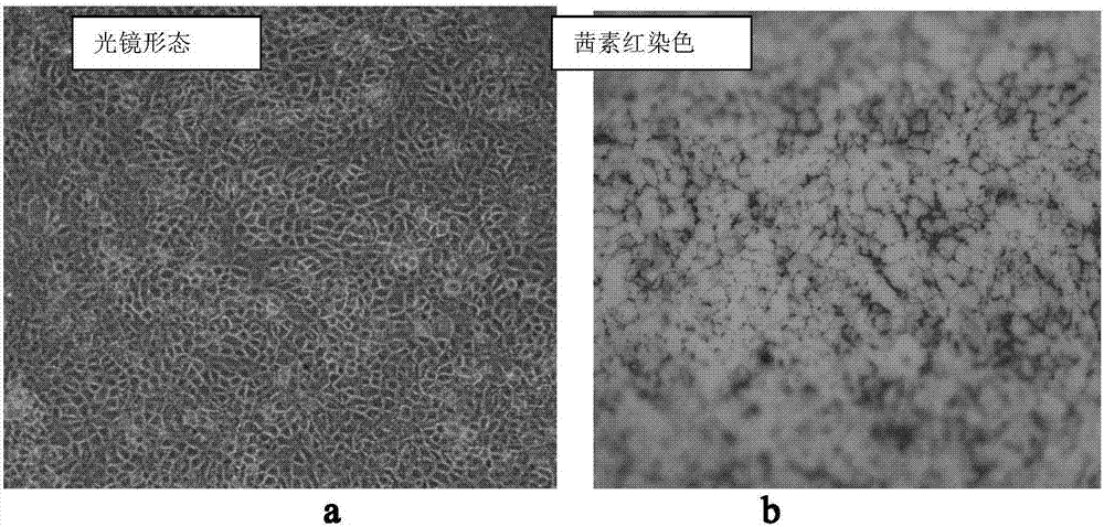
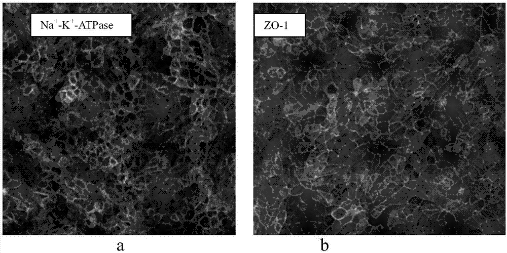
[0029] 1. Preparation of porcine corneal posterior elastic layer: take fresh whole porcine cornea, soak and rinse in sterile normal saline containing blue chain double antibody (blue chain double antibody (100×, purchased from Corning, US)) for 20 minutes. Place the face up under a stereo microscope, use a cotton swab to carefully remove the porcine corneal endothelial cells, use toothed forceps to fix the porcine cornea, first tear off the residual iris root and expose the free part of the elastic layer, and then use flat-head tweezers to carefully grasp the pig When the posterior elastic layer is free, gently tear to the center of the cornea to completely separate the pig posterior elastic layer from the corneal stroma. Pay attention to the operating strength to avoid tearing of the back elastic layer.

[0030] Put the completely separated porcine posterior elastic layer flat on a sterile rubber ring with an inner diameter of 9.0mm, and attach it to the center of a 35mm culture...


PUM


Abstract

The invention provides a method for establishing tissue engineering corneal endothelium. The method comprises the following steps: preparing a descemet membrane of swine corneal, culturing and inducing human multipotent stem cells, establishing tissue engineering corneal endothelium, and the like. Compared with an amniotic membrane or other carriers, the tissue engineering corneal endothelium established by using the method provided by the invention is not too soft and is good in transparency as the descemet membrane self is a normal cornea structure. Seed cells used in the method are induced and differentiated from the human multipotent stem cells, and are not limited by the multiplication capacity of primary cells. In addition, the tissue engineering corneal endothelium provided by the invention is simple in preparation process, wide in source and easy to operate.

Description

Technical field [0001] The invention belongs to the field of tissue engineering, and specifically relates to a method for constructing a tissue engineering corneal endothelium. Background technique [0002] The corneal endothelium is a single layer of cells attached to the Descemet's membrane and located on the innermost side of the cornea. It directly contacts the aqueous humor and maintains the cornea's transparency and normal thickness through its active fluid pump function. Adult corneal endothelial cells cannot regenerate. After damage, they are mainly repaired by the expansion and migration of cells around the damaged area. Various reasons such as Fuch corneal endothelial dystrophy, trauma and surgery can cause corneal endothelial cell damage, when the cell density is less than 500-1500 cells / mm 2 Decompensation of corneal endothelial function occurs from time to time, leading to corneal edema and turbidity, showing bullous keratopathy. For this type of disease, it can be ...

Claims


Application Information

Patent Timeline
Patent Type & Authority Applications(China)
IPC IPC(8): A61L27/36A61L27/38C12N5/071
CPCA61L27/3633A61L27/3687A61L27/3691A61L27/3834A61L2430/16C12N5/0697C12N2506/02C12N2506/1346C12N2506/45C12N2533/90
Inventor 段豪云周庆军史伟云李文静
Owner SHANDONG EYE INST
Why Eureka
Try Eureka