X-ray genetic marker probe based on synchrotron light source and preparing method and application thereof

A technology of genetic labeling and synchronizing light source, applied in the field of biochemistry, can solve the problems of inability to recognize and image intracellular biomolecules with high specificity, and achieve the effect of good specificity and good specificity

Active Publication Date: 2017-08-11
SHANGHAI INST OF APPLIED PHYSICS - CHINESE ACAD OF SCI
View PDF2 Cites 5 Cited by
  • Summary
  • Abstract
  • Description
  • Claims
  • Application Information

AI Technical Summary

Problems solved by technology

[0004] The purpose of the present invention is to provide an X-ray genetic marker probe based on a synchronous light source and its preparation method and application, so as

Method used


Image

  • X-ray genetic marker probe based on synchrotron light source and preparing method and application thereof
  • X-ray genetic marker probe based on synchrotron light source and preparing method and application thereof
  • X-ray genetic marker probe based on synchrotron light source and preparing method and application thereof

Examples


Embodiment 1

[0033] Example 1 Preparation of Synchronous X-ray Genetic Labeling Probe and Its Application in Cellular Connexin Imaging

[0034] Construction of pcDNA3-Cx43-APEX2 plasmid. The construction process of the plasmid was carried out by conventional molecular biology methods, as follows: First, according to the human Cx43 protein sequence, its DNA sequence was optimized using human-biased codons and the complete sequence was synthesized. The complete sequence was provided by Shanghai Quanyang Synthesized by Biotech Ltd. The APEX2 sequence was cloned from the pEGFP-APEX2-Tubulin plasmid (Addgene plasmid #66171). A linker sequence was connected between the Cx43 sequence and the APEX2 sequence using the Q5 site-directed mutagenesis kit (purchased from NEB, catalog number E0552S). The Cx43-APEX2 sequence was then cloned into the backbone of the pcDNA3 mammalian expression vector to construct the pcDNA3-Cx43-APEX2 plasmid. Sequence and verify the plasmid sequence. The plasmid seque...

Embodiment 2

[0039] Example 2 Preparation of Synchronous X-ray Genetic Labeling Probe and Its Application in Intracellular Tubulin Imaging

[0040] The pEGFP-APEX2-Tubulin plasmid (Addgene plasmid #66171) was purchased from Addgene. The plasmid sequence is shown in SEQ ID NO:2.

[0041] Culture of HeLa cells. Among them, HeLa cells were purchased from the Cell Bank of the Type Culture Collection Committee of the Chinese Academy of Sciences. MEM (containing 10% FBS) medium, 37°C, 5% CO 2 , cultured in saturated humidity. Put the silicon nitride window in the cell culture plate, sterilized by ultraviolet light, 8×10 4 Cells / well density were seeded and allowed to adhere overnight.

[0042] The pEGFP-APEX2-Tubulin plasmid was transfected into HeLa cells. The liposome Lipo3000 method was used for transfection, and 0.75 μL Lipo3000, 500 ng pcDNA3-Cx43-APEX2 plasmid and 1 μL P3000 were added to each well. After 24 hours, the medium was removed, and 2% glutaraldehyde was fixed in an ice bat...

Embodiment 3

[0044] Example 3 Preparation of Electron Microscopy Genetic Probe and Application Comparison of X-ray Genetic Probe in Simultaneous X-ray Cell Imaging

[0045] The pcDNA3-Cx43-APEX2 plasmid was transfected into HEK293T cells. Wherein, HEK293T cell culture and transfection method of pcDNA3-Cx43-APEX2 plasmid are the same as in Example 1.

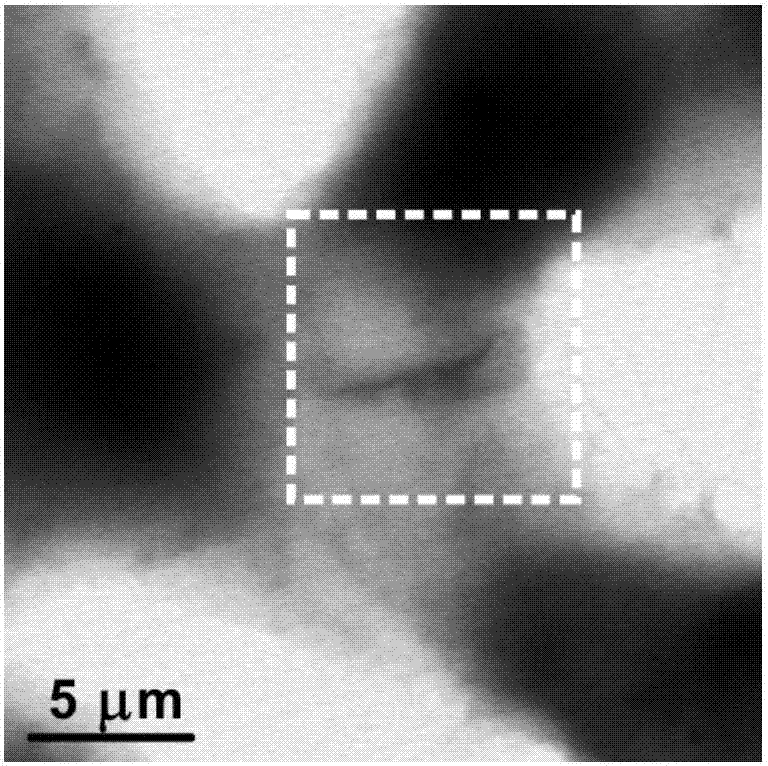
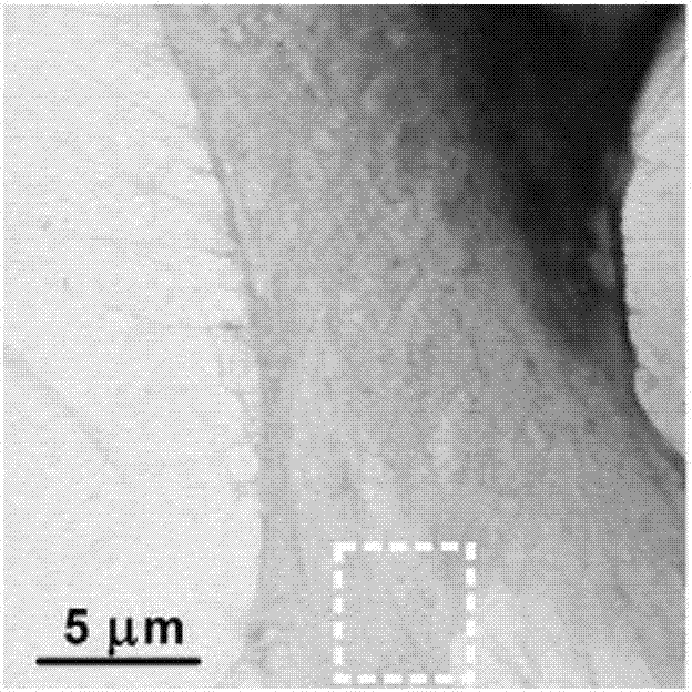
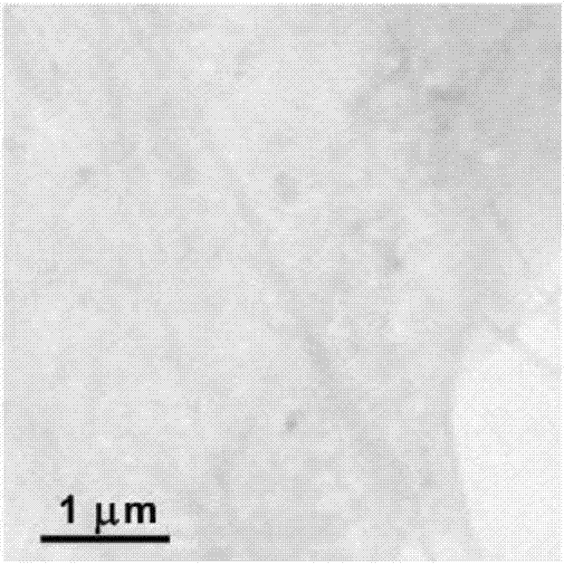
[0046] The labeling method of the electron microscope genetic probe is as follows: after the transfection, the culture medium in the well is removed, and the cells are fixed in an ice bath with 2% glutaraldehyde. Add containing 0.03% H 2 o 2 DAB reaction solution, react in ice bath for 1 min. Remove the reaction solution in the well, add 2% osmic acid, and counterstain in ice bath for 1 h. The reaction solution in the well was removed, dehydrated with gradient ethanol, and observed by X-ray imaging simultaneously, the method was the same as in Example 1.

[0047] The labeling method of the X-ray genetic probe and the X-ray imaging observ...


PUM


Abstract

The invention provides a method for preparing an X-ray genetic marker probe based on a synchrotron light source. The preparing method comprises the steps of 1, constructing fusion expression plasmids containing enzyme and target protein at the same time, and transfecting the fusion expression plasmids into cells; 2, conducting ice-bathing fixation of the cells by means of glutaraldehyde fixing liquid; 3, adding substrate molecule reaction liquid for ice-bathing reaction; 4, removing the reaction liquid, and fixing the cells with the fixing liquid; and 5, conducting synchrotron X-ray imaging observation, wherein the enzyme has substrate molecule catalytic activity. The invention further provides an X-ray genetic marker probe based on a synchrotron light source prepared with the preparing method, and application of the probe to cell imaging. The method can be used for high-specificity recognition and high-resolution imaging of intracellular biomolecules, and has broad biomedicine application prospects.

Description

technical field [0001] The invention relates to the technical field of biochemistry, and more specifically relates to an X-ray genetic marker probe based on a synchronous light source, a preparation method and application thereof. Background technique [0002] Microscopic imaging technology is one of the main driving forces for the development of cell life science. Especially in the past few decades, the emergence of fluorescence microscopy combined with labeling technology has realized the tracing of various biological macromolecules in cells, providing a revolutionary means for exploring various life activities of cells. However, subcellular structures and various biomacromolecules in cells are mostly in the 10-100nm scale. Due to the existence of the Abbe (Ernst Abbe) optical diffraction limit (200-300nm), it is difficult for traditional fluorescence microscopy techniques to detect particles below 100nm. Imaging observation of cell ultrastructure and important intracellu...

Claims


Application Information

Patent Timeline
IPC IPC(8): C12Q1/02G01N23/04
CPCG01N23/04G01N33/5005G01N2223/04
Inventor 樊春海诸颖孔华庭张继超夏凯王丽华胡钧
Owner SHANGHAI INST OF APPLIED PHYSICS - CHINESE ACAD OF SCI
Why Eureka
Try Eureka