Immune adsorption material for fixed point immobilizing protein A and preparation method thereof

An immunoadsorption material and protein technology, applied in the direction of solid adsorbent liquid separation, separation methods, chemical instruments and methods, etc., can solve the problems of reduced adsorption capacity, low antibody adsorption capacity, and inability to remove pathogenic antibodies by one-time perfusion , to improve the adsorption performance

Inactive Publication Date: 2017-08-18
重庆希尔康血液净化器材研发有限公司
View PDF6 Cites 8 Cited by
  • Summary
  • Abstract
  • Description
  • Claims
  • Application Information

AI Technical Summary

Problems solved by technology

Since there are many amino groups on the surface of protein A, which are distributed in different parts of protein A, protein A will be randomly coupled to the carrier in different parts, which cannot ensure that the active center of protein A is fully exposed, reducing its adsorption capacity. The adsorption capacity of the adsorption material for antibodies is about 20mg / mL
Because the adsorption capacity of the adsorption column for antibodies is not high, the purpose of eliminating pathogenic antibodies cannot be achieved by one-time perfusion, which hinders the wide application of protein A immunoadsorption therapy in clinical practice.

Method used


Image

  • Immune adsorption material for fixed point immobilizing protein A and preparation method thereof
  • Immune adsorption material for fixed point immobilizing protein A and preparation method thereof
  • Immune adsorption material for fixed point immobilizing protein A and preparation method thereof

Examples


Embodiment 1

[0029] Example 1: Preparation of recombinant protein A with cysteine

[0030] The coding sequence of the recombinant protein A with cysteine ​​is obtained by artificial synthesis, and the sequence connects the B domains in the protein A coding sequence (SEQ.ID.NO: 1) to form 5 B domains Concatenate, the recognition sequence of restriction endonuclease NdeI located at the 5' end of the sequence; a codon tgt encoding cysteine ​​and 6 histidine codons caccaccaccaccaccac located at the 3' end of the sequence XhoI recognition sequence.

[0031] SEQ.ID.NO.1

[0032]CATATGGTAGACaacaaattcaacaaagaacaacaaaatgctttctatgaaatcttacatttacctaacttaaacgaagaacaacgcaatggtttcatccaaagcctGaaagatgacccaagccaaagcgctaaccttttagcagaagctaaaaagctaaatgatgcGcaagcaccaaaaGTAGACaacaaattcaacaaagaacaacaaaatgctttctatgaaatcttacatttacctaacttaaacgaagaacaacgcaatggtttcatccaaagcctGaaagatgacccaagccaaagcgctaaccttttagcagaagctaaaaagctaaatgatgcGcaagcaccaaaaGTAGACaacaaattcaacaaagaacaacaaaatgctttctatgaaatcttacatttacctaacttaaacga...

Embodiment 2

[0035] Example 2: Preparation of Immunoadsorbent Material

[0036] Activation of Sepharose

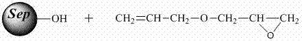
[0037] Add 60 ml of 2.5 M sodium hydroxide aqueous solution containing 0.2% sodium borohydride to 30 ml of drained agarose gel Sepharose 6FF, mix well, shake in a shaker (50-200rpm) for 2 hours, and the reaction temperature is 40°C, and then add 30ml of allyl glycidyl ether to continue the reaction for 6 hours. After the reaction is complete, rinse thoroughly with distilled water and drain to obtain agarose gel with alkenyl groups.

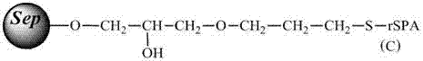
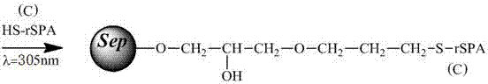
[0038] Immobilization of protein A

[0039] Dissolve 210 mg of protein A in Example 1 in 30 ml of 0.1 mol / mL phosphate buffer solution (pH=7.4), and then add it to 30 ml of the above-mentioned activated agarose gel (that is, each ml of agarose gel corresponds to 7 mg of protein A), under the irradiation of ultraviolet light with a wavelength of 305nm, shake at room temperature (50-200rpm) for about 10 hours, recover the unreacted protein A solution, was...

Embodiment 3

[0040] Embodiment three: the preparation of adsorption material

[0041] Activation of Sepharose

[0042] Operate in the same way as in Example 2 to obtain agarose gel with alkenyl groups.

[0043] Immobilization of protein A

[0044] Dissolve 180 mg of protein A in Example 1 in 30 ml of 0.1 mol / mL phosphate buffer solution (pH=7.4), and then add it to 30 ml of the above-mentioned activated agarose gel (that is, each ml of agarose gel corresponds to 6 mg of protein A), under the irradiation of ultraviolet light with a wavelength of 305nm, shake at room temperature (50-200rpm) for about 16 hours, recover the unreacted protein A solution, wash the adsorbent with water several times, and drain it. Add 30ml of PBS solution containing 0.2M mercaptoethanol, shake at room temperature (50-200rpm) for 4 hours, wash with distilled water for several times, and drain to obtain an adsorption material for immobilizing protein A. The fixed amount of protein A detected by biuret method was...


PUM

PropertyMeasurementUnit
adsorptionaaaaaaaaaa

Abstract

The invention belongs to the field of biological separation engineering and relates to a protein A immune adsorption material used for purifying blood and a preparation method thereof. The invention discloses a biopolymer separating material coupled with agarose gel microspheres and recombinant protein A. An immune adsorption material with high protein A absorption performance is prepared according to the following steps: taking agarose gel as a carrier, activating with allyl glycidyl ether, coupling with sulfydryl on recombinant protein A amino terminal (C terminal) cysteine, and finally, blocking the terminal, thereby fixing the C terminal of the protein A on the agarose gel microspheres at a fixed point. The material has the advantages that the recombinant protein A on the agarose gel microspheres is uniform in structure and fixed in sequence, so that the adsorption capacity of the adsorption material for an antibody is promoted, and the adsorption material can be used for clinical immune adsorption treatment.

Description

technical field [0001] The invention belongs to medical biomaterials, and in particular relates to a protein A immunoadsorbing material used for blood purification and a preparation method thereof. Background technique [0002] Even with the advancement of medicine today, there are still many intractable diseases that cannot be overcome by humans, and intractable autoimmune diseases are one of them. The pathogenesis of autoimmune diseases is complex and can affect multiple organs and systems of the body. The traditional treatment options are mainly glucocorticoids and cytotoxic drugs, but the therapeutic effect has been poor. It is currently the main cause that is difficult to deal with and threatens human health and life. one of the diseases. [0003] Immunoadsorption therapy is a new technology developed in the past ten years. It combines substances with specific affinity for pathogenic antibodies as ligands and carriers to prepare adsorption columns to selectively remove...

Claims


Application Information

Patent Timeline
Patent Type & Authority Applications(China)
IPC IPC(8): B01J20/24B01J20/30B01D15/38
CPCB01D15/3809B01J20/24
Inventor 罗章凯张文
Owner 重庆希尔康血液净化器材研发有限公司
Why Eureka
Try Eureka