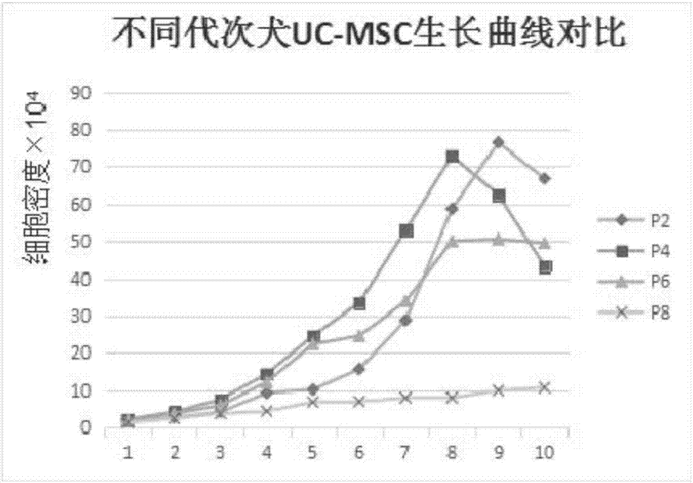
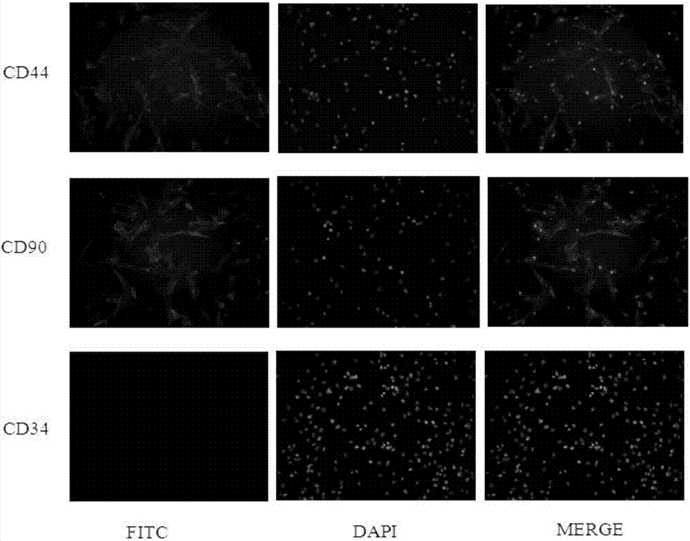
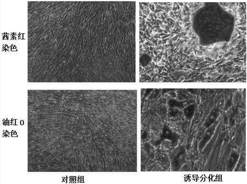
Method for performing primary culture on canine umbilical cord-derived mesenchymal stem cells

A technology of stromal stem cells and primary culture, applied in the field of bioengineering, can solve the problems of few reports on canine umbilical cord mesenchymal stem cells and the lack of effective treatment drugs for diseases. Effect

Active Publication Date: 2017-09-15
维赛(佛山)生物科技有限公司
View PDF1 Cites 14 Cited by
  • Summary
  • Abstract
  • Description
  • Claims
  • Application Information

AI Technical Summary

Problems solved by technology

Dogs are increasingly popular as pets, and the treatment technology for their diseases is developing rapidly. However, there are still no effective drugs for diseases such as arthritis, nerve damage and kidney damage. The treatment of mesenchymal stem cells provides a new way for this. train of thought
[0003] At present, the research on human mesenchymal stem cells is relatively in-depth, but there are few reports on the research on canine umbilical cord mesenchymal stem cells

Method used


Image

  • Method for performing primary culture on canine umbilical cord-derived mesenchymal stem cells
  • Method for performing primary culture on canine umbilical cord-derived mesenchymal stem cells
  • Method for performing primary culture on canine umbilical cord-derived mesenchymal stem cells

Examples


Embodiment 1

[0027] Embodiment 1: (collagenase method)

[0028] 1) Collection of newborn dog umbilical cord:

[0029] When the puppies are just born from the mother, use sterile instruments to strip the placenta, cut the umbilical cord, and immediately put the umbilical cord into the PBS buffer solution containing 10% double antibody solution;

[0030] 2) Isolate glial tissue rich in mesenchymal stem cells from the umbilical cord:

[0031] Cut the umbilical cord capsule to expose the tubular structure in the middle, remove the artery and vein, and the remaining hollow milky white tube without blood is the required tubular tissue;

[0032] 3) Cleaning and shredding the tissue pieces:

[0033] (3-1). Soak the isolated tubular tissue in PBS buffer solution containing 10% double antibody for 5 minutes, and take it out with tweezers;

[0034] (3-2). Soak the tubular tissue taken out in step (3-1) in PBS buffer solution containing 5% double antibody for 5 minutes, and take it out with tweezer...

Embodiment 2

[0048] Embodiment 2: (trypsin method)

[0049] This implementation is one of control examples, and the difference between its culture method and embodiment 1 is:

[0050] Step 4) Add about 5 times the volume of 0.25% trypsin, digest in a 37°C water bath for 30 minutes, and shake it from time to time to fully react the digestive juice with the tissue block.

[0051] The trypsin was purchased from Hyclone Company.

Embodiment 3

[0052] Embodiment 3: (tissue block method)

[0053] This implementation is used as one of the control examples, and the difference between its culture method and Example 1 is that steps 4) and 5):

[0054] 4-1) Evenly spread the cut umbilical cord tissue pieces on the bottom of a T25 culture bottle with the cut side down, and then place the bottom side up in 5% CO 2 , Cultivate in a 37°C incubator for 30 minutes;

[0055] 4-2) Take out the culture bottle, add 1ml of umbilical cord mesenchymal stem cell culture medium (just below the bottom of the bottle, to avoid suspending the tissue pieces), place the bottom side down in 5% CO 2 , Cultivate in a 37°C incubator for 2 hours;

[0056] 4-3) Take out the culture bottle, add 2ml of umbilical cord mesenchymal stem cell culture medium, completely submerge the tissue block, place the bottom side down in 5% CO 2 , Cultured in a 37°C incubator.


PUM


Abstract

The invention provides a method for performing primary culture on canine umbilical cord-derived mesenchymal stem cells. The mesenchymal stem cells are derived from umbilical cords of neonatal dogs and are wide in source and readily available in materials, so that large-scale production and clinical application of the cells become possible in the later period. The culture method is easy to operate and high in repeatability, and a few parenchyma cells and pollution-free pure mesenchymal stem cells can be obtained. A collagenase digestion method used in the culture method is mild, cell damage is less, and lots of mesenchymal stem cells with high reproductive capacity can be obtained and can be used for subsequent cell line establishment, characteristics of stem cells, disease treatment and the like.

Description

technical field [0001] The invention relates to the technical field of bioengineering, in particular to a method for primary culture of canine umbilical cord-derived mesenchymal stem cells. Background technique [0002] Mesenchymal Stem Cells (MSCs) are a type of adult stem cells with multi-directional differentiation ability, which can be directed to differentiate into various cells such as muscle, cartilage, blood vessels, endothelial cells and nerves under specific conditions. A type of adult stem cells with application prospects. Many studies have proved that they have significant effects in the treatment of cardiovascular diseases, blood diseases, joint injuries and other diseases. Umbilical cord-derived mesenchymal stem cells have the advantages of easy-to-obtain materials and low immunogenicity, and avoid ethical and moral disputes in disease treatment, so they have broad market prospects. Dogs are increasingly popular as pets, and the treatment technology for their ...

Claims


Application Information

Patent Timeline
Patent Type & Authority Applications(China)
IPC IPC(8): C12N5/0775
CPCC12N5/0665C12N2500/32C12N2500/84C12N2509/00
Inventor 王丙云詹小舒詹贝莹陈志胜计慧琴陈胜锋
Owner 维赛(佛山)生物科技有限公司
Why Eureka
Try Eureka