Double-sided hollow nanoneedle array device and preparation method thereof

A technology of hollow nano and needle arrays, which is applied in the field of intercellular protein transport, can solve problems affecting cell life activities, time-consuming and cumbersome processes, and cytotoxicity, so as to treat abnormal cell protein deficiency, maintain integrity and activity, and avoid The effect of normal function

Active Publication Date: 2019-07-30
SUN YAT SEN UNIV
View PDF1 Cites 0 Cited by
  • Summary
  • Abstract
  • Description
  • Claims
  • Application Information

AI Technical Summary

Problems solved by technology

[0008] First, the hybrid cells obtained after the cell fusion method have chromosomal aneuploidy, which causes the genetic instability of the cell line, and cannot selectively make the recipient cells obtain the required proteins, but absorbs many other proteins of the donor cells together, The medium required in the fusion process is cytotoxic, causing great damage to the cells and affecting the life activities of the cells, and the donor cells cannot provide protein again after fusion
[0009] Second, the disadvantage of the DNA transfection method is that the steps for preparing the DNA transfection plasmid are cumbersome. It is necessary to transfect the DNA in the cell to bacteria first, so that the bacteria can express a large amount of DNA transfection plasmid, and then further extract and purify to obtain the DNA plasmid
The particle size of DNA nanocomplexes is unstable, and in order to obtain higher transfection efficiency, a higher DNA concentration is often required, and a higher carrier / DNA complex concentration often causes certain cytotoxicity and affects the normal life activities of cells
[0010] Third, the direct protein transfer method needs to extract the protein from the donor cell through cell lysis or through a microneedle device, then separate and purify it, and then transport it to the abnormal cell. The process may cause protein contamination or denaturation, and it is easy to cause greater damage to cells without minimally invasive extraction and delivery devices
At present, there is still a lack of an effective technology that can simultaneously extract and deliver a specific protein from a batch of donor cells to recipient cells non-destructively

Method used


Image

  • Double-sided hollow nanoneedle array device and preparation method thereof

Examples


preparation example Construction

[0042] The steps for preparing the upper hollow nanoneedle array or the lower hollow nanoneedle array described in the above step (1) are as follows:

[0043] Use a polycarbonate substrate membrane with uniform nanopore size as a template

[0044] a. First, use atomic layer deposition technology, using gas phase tris or dimethylamino or silane as a precursor and water vapor pulses to alternately pass into the reactor, and deposit uniform silicon oxide on all surfaces of the template substrate including inner pore walls Floor;

[0045] b. Then use plasma etching method, use SF 6 and CF 4 The gas etches away the silicon oxide on the upper surface;

[0046] c, then further use O 2 Part of the substrate film is etched away by plasma etching to form a tubular hollow nanoneedle structure of silicon oxide.

[0047] The steps of integrating the hollow nanoneedle array described in the above step (2) with the microfluidic pipeline are as follows:

[0048] a, use photolithography ...

Embodiment 1

[0059] Transport of GFP between Hela cells:

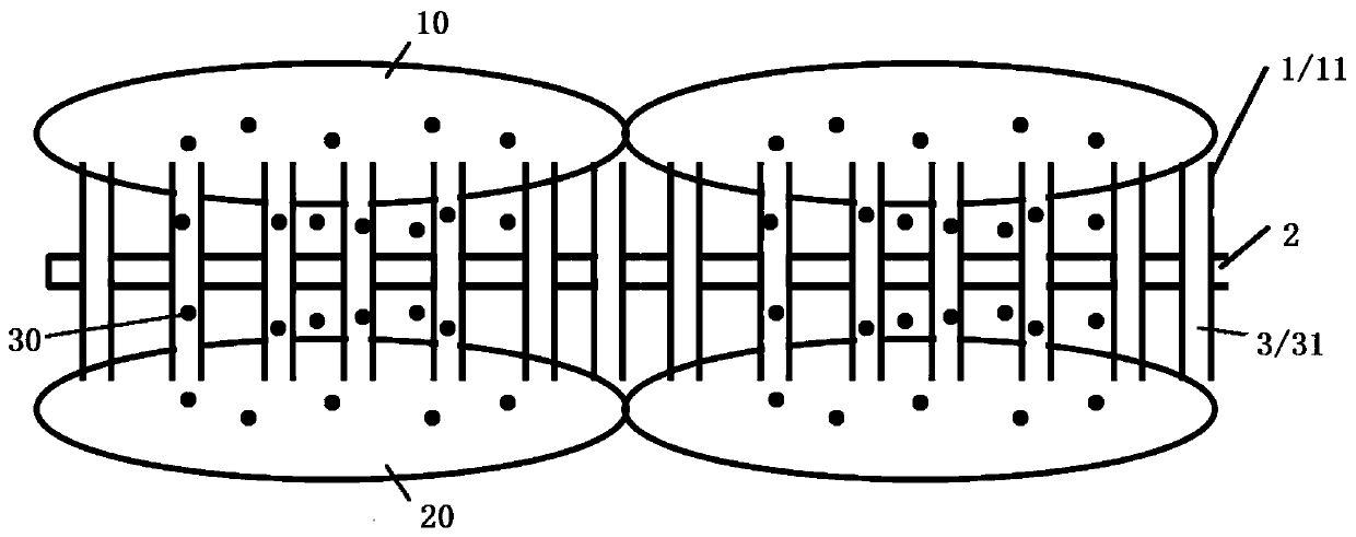
[0060] A number of Hela cells are placed on the upper layer of the double-sided nanoneedle array device, and the mutant Hela cells with green fluorescent protein are placed on the lower layer to provide an environment suitable for the normal life of the cells. Flowing into the microfluidic pipeline 2, the green fluorescent protein 30 in the lower cell 20 flows into the upper cell 10, and the green fluorescent protein in the upper and lower Hela cells of the same batch is observed with a confocal fluorescence microscope every 12 hours. It was found that most of the Hela cells were still living normally after repeated extractions, and green fluorescent protein was detected in the Hela cells without green fluorescent protein. Intracellular protein is extracted creatively and transported to another type of cell to maintain the integrity and activity of the cell and avoid interfering with the normal function of the cell; it can continuo...

Embodiment 2

[0062] Transport of green and red fluorescent proteins between Hela cells:

[0063] A number of Hela cells expressing green fluorescent protein are placed on the upper layer of the double-sided nanoneedle array device, and mutant Hela cells with red fluorescent protein are placed on the lower layer to provide an environment suitable for the normal life of the cells, and the hollow nanoneedle 11 on the upper layer is used to break the Hela cell membrane , so that the cell liquid flows into the microfluidic channel 2 , and the red fluorescent protein 30 in the lower layer cells 20 flows into the upper layer cells 10 . The red and green fluorescent proteins in the upper and lower Hela cells of the same batch were observed with a confocal fluorescence microscope every 12 hours. It can be found that most of the Hela cells are still living normally after repeated extractions, and red and green fluorescent proteins are detected in the upper and lower Hela cells at the same time. Int...


PUM

PropertyMeasurementUnit
pore sizeaaaaaaaaaa

Abstract

The invention belongs to the technical field of intercellular protein transmission, and concretely discloses a two-sided hollow nanoneedle array device. The two-sided hollow nanoneedle array device comprises hollow nanoneedle head arrays which are arranged at the upper layer and the lower layer and are formed by arranging a plurality of hollow nanoneedle heads; all hollow nanoneedle heads of the upper and lower hollow nanoneedle head arrays are corresponding; each hollow nanoneedle head of the upper hollow nanoneedle head array is communicated with a micro-fluid channel; each hollow nanoneedle head of the lower hollow nanoneedle head array is also communicated with the micro-fluid channel; the two end parts of the micro-fluid channel are provided with small holes. The array device has the advantages that on the premise of not destroying the cell activity, the specific protein in a large number of donor cells can be extracted in a minimally invasive mode and is conveyed into recipient cells; the completeness and the activity of the cells are maintained; the interference with the normal functions of the cells is avoided; the protein can be continuously provided for the recipient cells to achieve the goal of cell treatment.

Description

technical field [0001] The invention belongs to the technical field of intercellular protein transmission, in particular to a double-sided hollow nanoneedle array device and a preparation method thereof. Background technique [0002] With the development of biomedical technology, there are many ways to study the transport of substances between cells. Protein molecules in cells are one of the main research objects in the field of bioelectronics and bioinformatics. They play a key role in regulating cell activities and maintaining cell or body functions. The loss of any protein molecule in the cell will have a great impact on the normal life activities of the cell, so the study will send the missing protein to the abnormal cell and maintain the activity of the donor cell The method has great significance for biology and medicine. [0003] At present, a series of mature technologies are used to supplement missing proteins to repair cell defects, such as cell fusion, DNA trans...

Claims


Application Information

Patent Timeline
Patent Type & Authority Patents(China)
IPC IPC(8): C07K1/14
CPCC07K1/145
Inventor 谢曦潘烁琳柳成林肖帅林迪安陈惠琄杭天杨成端吴江明辜美霖李恩来
Owner SUN YAT SEN UNIV
Why Eureka
Try Eureka