CD4+T cell derived exosomes and application thereof

A cell and source technology, applied in the direction of blood/immune system cells, animal cells, vertebrate cells, etc., to achieve the effect of strengthening humoral immune response, fast speed and short time consumption

Active Publication Date: 2017-11-21
JIANGSU UNIV
View PDF1 Cites 3 Cited by
  • Summary
  • Abstract
  • Description
  • Claims
  • Application Information

AI Technical Summary

Problems solved by technology

Can the exosomes derived from it also play a role in promoting the body's immune response? Current studies have confirmed that M1-polarized macrophages can be used as immune enhancers f

Method used


Image

  • CD4+T cell derived exosomes and application thereof
  • CD4+T cell derived exosomes and application thereof
  • CD4+T cell derived exosomes and application thereof

Examples


Embodiment 1

[0039] Example 1: CD4 + Sorting of T cells and preparation of culture supernatant

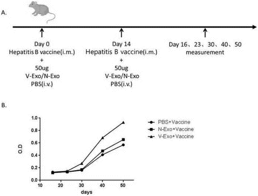
[0040] (1) Establish the immune mouse model: Babl / c mice (Animal Experiment Center of Jiangsu University) were divided into three groups for immunization.

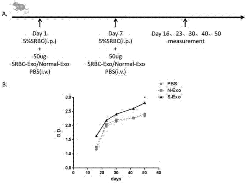
[0041] Sheep red blood cell (SRBC) mouse immunization: 5% SRBC was intraperitoneally injected into 6-8w female Babl / c mice (Animal Experiment Center of Jiangsu University), and then injected again 7 days later for a booster immunization. On the 9th day, the mice were sacrificed to separate spleen cells.

[0042] Ovalbumin (OVA) (Sigma) mice were immunized: 100µg OVA was dissolved in 100µLPBS, and the same amount of complete Freund's adjuvant was added to grind it into a water-in-oil state, and 6-8w female Babl / c mice were subcutaneously injected into the right back at multiple points, After 7 days, double OVA was fused in PBS, and multi-point subcutaneous booster immunization was performed on the contralateral back. On the 9th day, the mi...

Embodiment 2

[0047] Example 2: CD4 + Preparation of T Exo and determination of protein concentration

[0048] (1) The sorted CD4 + T cells according to 2×10 6 Cells / well were seeded in a 24-well plate pre-coated with CD3, resuspended in 1 mL of 10% FBS 1640 culture medium, added 2 µg / mL CD28 to stimulate for 24 hours, and the supernatant was collected. The collected CD4 + T cell supernatant was centrifuged at 4°C and 300g for 20min to collect the supernatant; passed through a 0.22µm filter to collect the filtrate; then transferred the filtrate to a MWCO 100 kDa ultrafiltration centrifuge tube (Millipore), centrifuged at 1500g for 30min, and collected the inner tube concentrate in.

[0049] (2) Extract CD4 with the exoQuick-TCTM exosome kit purchased from SBI + T exo: Mix the concentrated solution obtained in step (1) with exoQuick-TCTM exosome reagent at a volume ratio of 5:1, shake, and place at 4°C for more than 12 hours; centrifuge at 1000g for 30 minutes at 4°C, discard the sup...

Embodiment 3

[0051] Example 3: CD4 + Identification of T Exo

[0052] (1) Observation of CD4 by transmission electron microscope + T Exo form: Take 20µL CD4 + Add the T Exo suspension dropwise on the sample-loading copper grid with a diameter of 3 mm, and let it stand at room temperature for 2 minutes; gently blot the liquid with filter paper, drop 2% phosphotungstic acid solution with pH 6.8 on the copper grid, and negatively stain for 1 min; blot the filter paper dry The dye solution was dried under an incandescent lamp and observed under a transmission electron microscope. The result is as figure 2 As shown, under the transmission electron microscope, CD4 can be observed + T Exo is a round or oval biconcave disc-shaped microcapsule structure with a complete envelope, and the cavity contains low electron density components, and the particle size is mainly distributed in the range of 30-110nm.

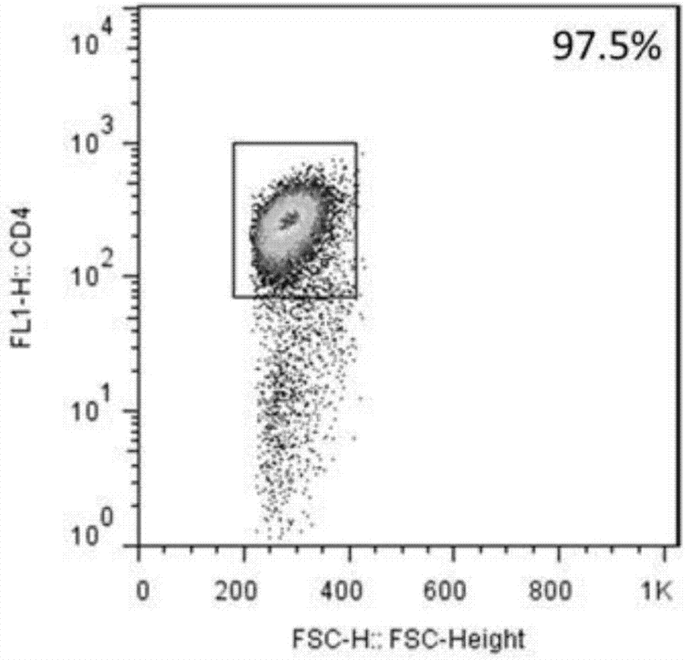
[0053] (2) Flow cytometric detection of exosomes surface molecules CD4 and CD25: add...


PUM


Abstract

The invention realtes to CD4+T cell derived exosomes and application thereof, and belongs to the field of cytobiology, molecular biology and clinical application. The invention particularly discloses CD4+T cell derived exosomes (named as CD4+ T exo) and application thereof in preparation an adjuvant for enhancing a vaccine immunity effect. It discovers that CD4+ T exo can effectively promote proliferation and activation of B cells in vitro, and can promote the B cells to produce more antibodies. Meanwhile, CD4+ T exo can serve as an adjuvant to enhance the immune effect of sheep red blood cell (SRBC) immunized mice in vivo, can also have obvious enhancing effect on hepatitis B vaccine immunized Babl/c mice, and promotes peripheral blood to produce more hepatitis B surface antibodies (HbsAb). The application of the CD4+T cell derived exosomes can be used for preparing a novel adjuvant for enhancing the antigen/vaccine immunization effect.

Description

technical field [0001] The present invention relates to a CD4 + Exosomes derived from T cells and their applications belong to the field of cell biology, molecular biology or clinical application, specifically related to CD4 isolated from mouse spleen + Preparation of T exo, study of its biological function and its application in strengthening the humoral immunity of mice. Background technique [0002] Exosomes are double-membrane vesicles with a diameter of 30-110 nm, which originate from early endosomes in cells and can be released into the extracellular environment after fusion with the plasma membrane and play biological roles. Studies have shown that almost all living cells can secrete exosomes, and they are widely present in various body fluids. Exosomes can carry protein, DNA, RNA and other components of the source cells, making them less susceptible to degradation by the external environment, which is conducive to the biological functions of related active compon...

Claims


Application Information

Patent Timeline
IPC IPC(8): C12N5/0783A61K39/39A61K39/29A61P1/16A61P31/20
CPCA61K39/12A61K39/39A61K2039/55555A61K2039/575C12N5/0636C12N2730/10134
Inventor 王胜军陆健田洁吴静王运刚马洁马斌许化溪
Owner JIANGSU UNIV
Why Eureka
Try Eureka