
Liquid-phase surface-enhanced Raman spectroscopy sensor, its preparation method and its use for nucleic acid detection

A surface-enhanced Raman and spectral sensor technology, applied in the field of functional nanomaterials and biological detection, can solve rare and other problems

Active Publication Date: 2020-07-07
元朗生命科技(南京)有限公司
View PDF4 Cites 0 Cited by
  • Summary
  • Abstract
  • Description
  • Claims
  • Application Information

AI Technical Summary

Problems solved by technology

However, most of the reported magnetic core-shell structures have smooth gold shell surfaces, but there are few studies on the growth of branched gold shells on the surface of magnetic cores to improve the SERS enhancement performance of particles.

Method used


Image

  • Liquid-phase surface-enhanced Raman spectroscopy sensor, its preparation method and its use for nucleic acid detection
  • Liquid-phase surface-enhanced Raman spectroscopy sensor, its preparation method and its use for nucleic acid detection
  • Liquid-phase surface-enhanced Raman spectroscopy sensor, its preparation method and its use for nucleic acid detection

Examples


Embodiment 1

[0040] Example 1 Preparation of magnetic core branched gold-shell nanoparticles:

[0041] (1) Prepare 20mM HAuCl 4 solution, the concentration is 0.1M K 2 CO 3 solution, concentration 10mM AgNO 3 solution.

[0042] (2) At room temperature, take the above 720 μL 0.1M K 2 CO 3 and 760 μL 20 mM HAuCl 4 Add 20mL of ultrapure water and stir vigorously for 20min to prepare the gold shell growth solution;

[0043] (3) 20μL 10mM AgNO 3 Add to 100 μL Fe 3 o 4 @SiO 2 @Au seeds and ultrasonic treatment for 10min;

[0044] (4) Mix the solutions obtained in steps (2) and (3), and stir vigorously for 2 minutes;

[0045] (5) Add 100 μL of formaldehyde solution to the mixed solution in step (4) and continue to stir for 2 minutes;

[0046] (6) After the mixture was left to grow for 30 minutes at 28 ° C, the synthesized colloid was separated and purified by a centrifuge at a speed of 2500 rpm, centrifuged for 15 minutes, washed with ultrapure water three times, and finally obtained...

Embodiment 2

[0047] Example 2 Preparation and detection method of nucleic acid detection SERS sensor

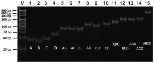
[0048] (1) The amount of four DNA single strands (A, B, C and D listed in Table 1) and other substances are mixed in TM buffer (20mM Tris-HCl, 50mM MgCl 2 , pH 8.0), and subjected to high-temperature cooling treatment, that is, kept in a constant temperature shaker at 95°C for 5min, and then kept in a refrigerator at 4°C for 20min to form a tetrahedral DNA probe with a final concentration of 1 μM. The formation of tetrahedral DNA probes was verified by 10% polyacrylamide gel electrophoresis. image 3 It is an electrophoretic gel image. Compared with the combination of one, two and three DNA strands, the tetrahedral DNA probe formed by the mixed self-assembly of four DNA strands moves the slowest in lane 15, which verifies the successful formation of the tetrahedral DNA probe. , and the yield is high.

[0049] (2) Mix 10 μL tetrahedral DNA solution with 500 μL magnetic core branched gold...


PUM

PropertyMeasurementUnit
diameteraaaaaaaaaa
lengthaaaaaaaaaa

Abstract

The invention discloses a liquid phase surface-enhanced Raman spectroscopy sensor, a preparation method thereof and application to nucleic acid detection. The liquid phase surface-enhanced Raman spectroscopy sensor comprises two parts, namely a detection substrate and an SERS probe, wherein the detection substrate consists of tetrahedral DNA probe-modified magnetic nuclear twig-like gold shell nanoparticles, and the SERS probe consists of gold nanoparticles of which the surfaces are modified with specific nucleotide sequences capable of being hybridized with a target nucleic acid as well as Raman signal molecules. During detection, the detection substrate, the SERS probe and a liquid sample to be detected are mixed, a detection substrate-target nucleic acid-SERS probe compound adopting a sandwich structure is formed through complementary base pairing, and the compound in a detection solution is separated by using an external magnetic field and is enriched for SERS testing; and high-sensitivity and specific detection of the nucleic acid in serum is achieved by virtue of an SERS signal, the detection limit is as high as fM, and detection of a nucleic acid marker in complex environments such as the serum can be achieved.

Description

technical field [0001] The invention belongs to the field of functional nanomaterials and biological detection, and in particular relates to a liquid-phase surface-enhanced Raman spectroscopy (SERS for short) sensor for nucleic acid detection, a preparation method thereof and a nucleic acid detection application thereof. Background technique [0002] With the deepening of research on the mechanism of tumorigenesis, people have gradually realized that tumor is essentially a disease caused by changes in genetic material. In recent years, studies have proved that the uncontrolled expression of miRNA is closely related to the occurrence and development of tumors. In specific tissues and specific development Stage expression has tissue specificity and timing, which determines the important role of miRNA in disease diagnosis. However, the content of tumor nucleic acid markers in the early stage of lesions is very low, and it is urgent to develop new methods for reliable, low-cost ...

Claims


Application Information

Patent Timeline
Patent Type & Authority Patents(China)
IPC IPC(8): G01N21/65C12Q1/6886
CPCC12Q1/6825G01N21/658
Inventor 宋春元汪联辉蒋新宇杨琰君窦艳霞孙友志董晨李放
Owner 元朗生命科技(南京)有限公司
Learn More