An ultra-low-field magnetic detection method for measuring cell adhesion and cell migration rate

A technology of cell migration and adhesion, applied in the field of biomedicine, can solve the problems of failing to achieve high throughput, and achieve the effect of wide application range and sensitive detection

Active Publication Date: 2020-07-31
INST OF CHEM CHINESE ACAD OF SCI
View PDF0 Cites 0 Cited by
  • Summary
  • Abstract
  • Description
  • Claims
  • Application Information

AI Technical Summary

Problems solved by technology

However, the obvious disadvantage of AFM technology is that it cannot achieve high-throughput and simultaneously measure the adhesion of a large number of cells.
Moreover, it is difficult for existing methods to use a certain platform to measure both the migration rate of cells and the adhesion of cells

Method used


Image

  • An ultra-low-field magnetic detection method for measuring cell adhesion and cell migration rate
  • An ultra-low-field magnetic detection method for measuring cell adhesion and cell migration rate
  • An ultra-low-field magnetic detection method for measuring cell adhesion and cell migration rate

Examples


Embodiment 1

[0055] The modification of the extracellular matrix protein FN of embodiment 1, FIRMS chip

[0056] Prepare a polydimethylsilane (PDMS) cuboid with a length, width and height of 1.4 cm, 0.6 cm and 0.2 cm respectively. In the middle of the cuboid is a circular depression with a diameter of 0.4 cm, wherein the concave part is the FIRMS detection part. Place the FIRMS chip with the concave side facing up in the Petri dish, and clean the chip with plasma for 40s. After the surface of the chip is activated, a 2% volume fraction of 3-aminopropyltriethoxysilane (APTES) absolute ethanol solution is quickly dropped. After reacting for 4 hours, the solution was discarded, and the chip was washed 5 times with absolute ethanol, and then washed 5 times with deionized water. Store in a desiccator after drying with nitrogen gas. Glutaraldehyde aqueous solution (1%, v / v) was added to the aminated chip, and after reacting for 3 hours, it was washed 5 times with deionized water and 5 times wi...

Embodiment 2

[0057] Embodiment 2, the magnetic labeling of cell

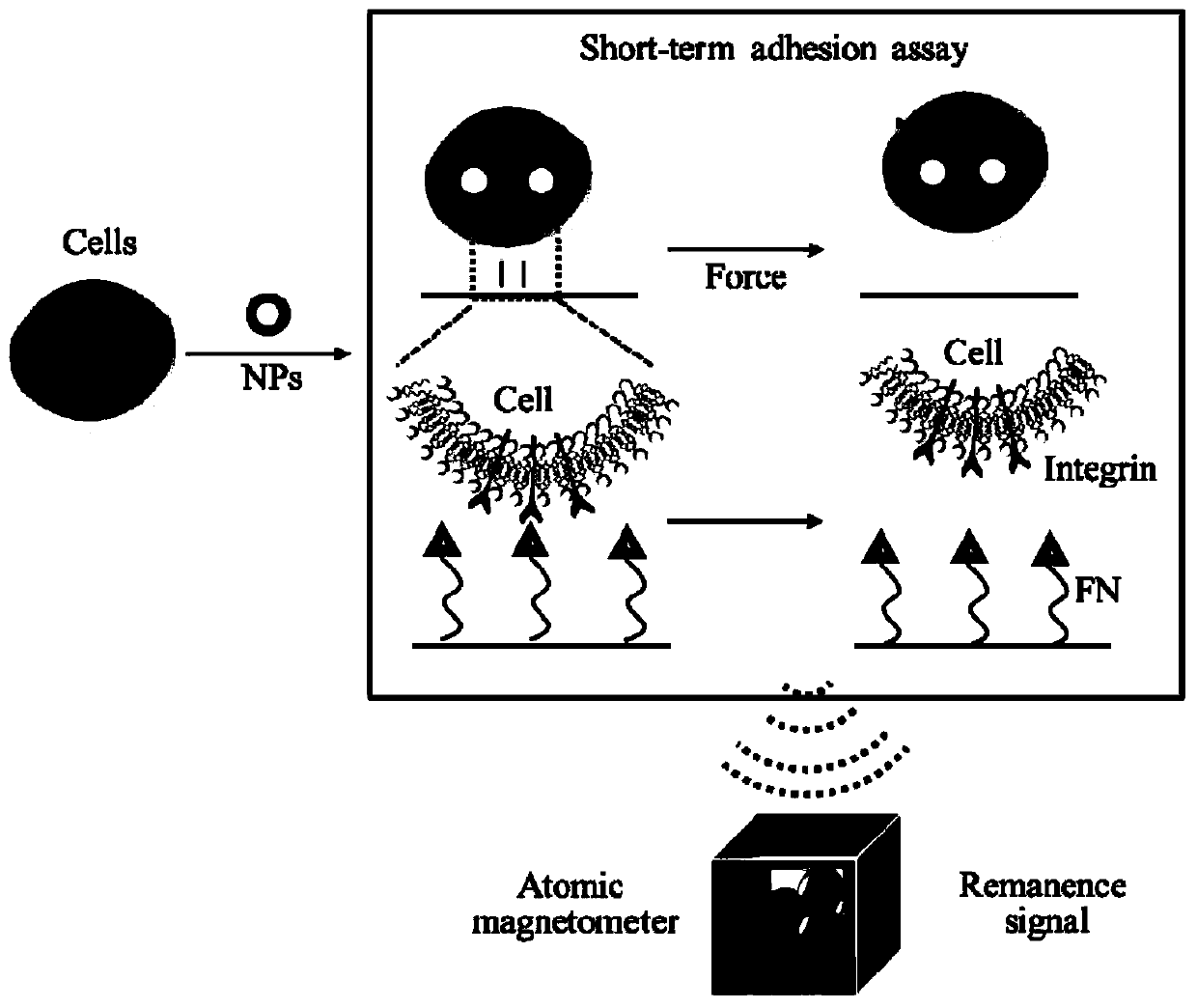
[0058] Will 1×10 6 Human lung cancer cells (A549) cells were inoculated in a 6-well cell culture plate, and after culturing for 17 hours, the cell culture medium was discarded. Cells were washed 3 times with serum-free culture. Serum-free medium dispersed with magnetic probes (20ug / mL) was added to continue culturing for 4h. The culture medium containing the magnetic probe was sucked off, and the cells were washed 5 times with phosphate buffer solution. The cells that had internalized the magnetic probe were then digested with trypsin (0.25%) for 3 min. After the digested cells were centrifuged to separate the solution containing trypsin, the cells were blown completely into the complete medium for later use.

Embodiment 3

[0059] Embodiment 3, FIRMS feasibility verification

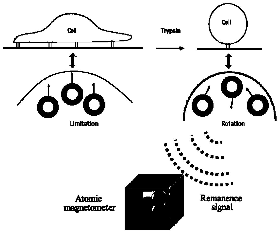
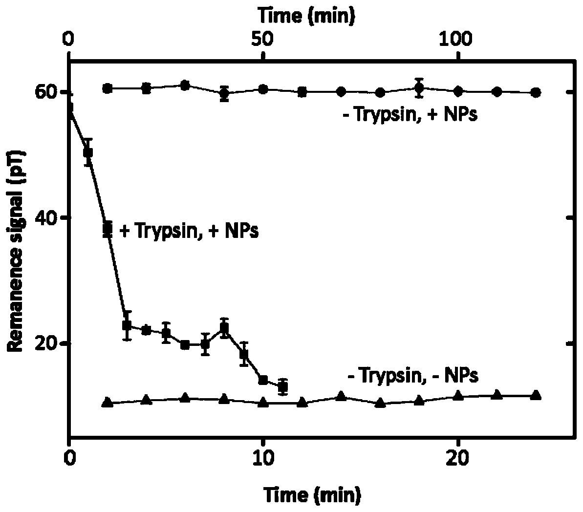
[0060] Such as figure 1 As shown, in order to verify that FIRMS can sensitively respond to external force interference or changes in the remanence signal caused by the cell's own migration, the cells were first cultured on the FIRMS chip. After culturing for 17 hours, the complete medium was discarded and washed with serum-free medium. The cells were added twice and incubated for 4 hours with the addition of a magnetic probe. After washing away the magnetic probes that are not endocytosed by the cells or adsorbed on the cell surface, the intracellular probes are magnetized for 2 min (the strength of the magnet is 1T). After adding 0.25% trypsin, quickly use FIRMS to record the contraction process of the cells under trypsinization, from the spreading state to the spherical shape until finally the decrease of the residual magnetic signal caused by the cells detaching from the substrate. Compared with the residual magnetic s...


PUM

PropertyMeasurementUnit
sizeaaaaaaaaaa
strengthaaaaaaaaaa
diameteraaaaaaaaaa

Abstract

The invention discloses an ultra-low field magnetic detection method for determination of cell adhesion and cell migration rate. The method includes the steps of: inoculating a magnetic labelled cellto a modified chip for incubation and magnetizing a magnetic probe in the magnetic labelled cell, then determining the cell adhesion and cell migration rate: 1) conducting external force interferenceof the magnetized cell and performing dissociation from the chip, at the same time using FIRMS to record the remanence signal drop spectrogram of the modified chip, and according to the spectrogram, defining the force corresponding to half of the remanence signal as the adhesive force for calculation, thus obtaining the cell adhesion; and 2) using FIRMS to record the remanence signal reduction spectrogram of the cell migration caused modified chip obtained by magnetization, and according to the spectrogram, conducting linear fitting calculation of remanence signal drop rate, thus obtaining thecell migration rate. The invention uses FIRMS to sensitively respond to the remanence signal change caused by external force or migration movement of the cell itself, and is not affected by distance.

Description

technical field [0001] The invention relates to an ultra-low field magnetic detection method for measuring cell adhesion and cell migration rate, belonging to the field of biomedicine. Background technique [0002] The migration of tumor cells is the most basic phenomenon in the process of tumor metastasis. Cell migration ability can reflect potential tumor metastasis. The complex process of cell migration is mainly driven by genes inside tumor cells as well as chemical factors outside tumor cells. These factors mainly include growth factors, cytokines and chemokines. In the past two decades, scientists have discovered that in addition to changes in genes and chemical factors within cells or tissues, physical interactions and mechanical forces between cells and cells, cells and their microenvironment play a very important role in the process of cell migration . Cell migration is currently described as a cyclic process coordinated in time and space by cytoskeletal protein...

Claims


Application Information

Patent Timeline
Patent Type & Authority Patents(China)
IPC IPC(8): C12Q1/02G01N27/72
Inventor 姚立张娣
Owner INST OF CHEM CHINESE ACAD OF SCI
Why Eureka
Try Eureka