

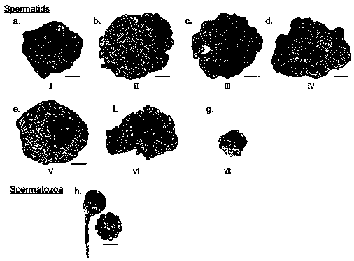
Cytology division method of scophthalmus maximus spermiogenesis phase
A technology of sperm cells and cytology, applied in the direction of scientific instruments, analytical materials, preparation of test samples, etc., can solve the problems of lack of system division, difficulty in observing and distinguishing the structure of sperm cells in different metamorphosis stages, limited, etc., and achieve high Reference and research value, improvement of dyeing uniformity, effect of enhancing image clarity
- Summary
- Abstract
- Description
- Claims
- Application Information
AI Technical Summary
Problems solved by technology
Method used
Image

Examples
Embodiment 1
[0029] The cytological division method of turbot sperm metamorphosis stage comprises the following steps:
[0030]1) Collect the testis of turbot in stage IV-V, put the testis tissue in 2.0% glutaraldehyde solution at 1°C, dissolve 25% glutaraldehyde in phosphate buffer solution to obtain glutaraldehyde solution, Fix at 4°C for 3 hours; rinse with phosphate buffer for 3 times, and post-fix with 1.1% osmic acid with pH 7.18 at 4°C for 1.0 hour; 2) Wash the fixed testis tissue with phosphate buffer for 3 The second time, take 0.4% uranyl acetate solution, soak and stain the testis tissue at 1°C overnight, and take it out with CO2-free 2 Wash off the excess dye solution in distilled water, then rinse with phosphate buffer three times for use; 3) Dehydrate with ethanol solutions with gradient concentrations of 50%, 70%, 80%, 90% and 95%, and treat each concentration for 15 minutes , and then treated with 100% alcohol for 20 minutes, then treated with pure ethylene oxide for 20 mi...
Embodiment 2
[0040] The cytological division method of the metamorphosis stage of turbot sperm cells includes: fixation, pre-staining, dehydration and embedding, uranyl acetate staining, lead citrate staining, and slice analysis, specifically including the following steps:
[0041] 1) Fixation: Collect turbot testis in stage IV-V, place testis tissue in 2.5% glutaraldehyde solution at 4°C, dissolve 25% glutaraldehyde in phosphate buffer solution to obtain glutaraldehyde solution, fixed at 5°C for 4 hours; washed with phosphate buffer for 4 times, and post-fixed with 1.2% osmic acid at 5°C for 1.3 hours; fixation with glutaraldehyde solution and osmic acid can make the substances in the sperm cells as much as possible The morphological structure and position when it is close to its living state can prevent the autolysis and corruption of sperm cells, prevent the enzymes in the cells from decomposing proteins, and turn various components in the cells such as proteins, fats, carbohydrates, etc...
Embodiment 3
[0057] The cytological division method of the metamorphosis stage of turbot sperm cells includes: fixation, pre-staining, dehydration and embedding, uranyl acetate staining, lead citrate staining, and slice analysis, specifically including the following steps:
[0058] Fixation: Collect turbot testis in stage IV-V, place testis tissue in 2.5% glutaraldehyde solution at 2°C, dissolve 25% glutaraldehyde solution in phosphate buffer solution to obtain 2.5% glutaraldehyde solution Dialdehyde solution, fixed at 4°C for 3.5 hours; rinsed with phosphate buffer three times, and then post-fixed with 1.2% osmic acid at 4°C for 1.0 hour; fixation with glutaraldehyde solution and osmic acid can make sperm cells The substance is as close as possible to its morphological structure and position in its living state, which can prevent the autolysis and corruption of sperm cells, prevent the enzymes in the cells from decomposing proteins, and make various components in the cells such as proteins...
PUM
 Abstract
Description
Claims
Application Information