Monoclonal antibody resistant to vole babesia and application thereof

A technology of Babesia vole and monoclonal antibody is applied in the field of blood protozoa control of parasitic diseases, which can solve the problems of low detection rate, high technical and experimental skills requirements of testing personnel, and difficulty in batch testing.

Active Publication Date: 2018-07-24
HUAZHONG AGRI UNIV
View PDF7 Cites 1 Cited by
  • Summary
  • Abstract
  • Description
  • Claims
  • Application Information

AI Technical Summary

Problems solved by technology

[0005] Blood smear staining microscopic examination is easy to operate and does not require high equipment, but it requires a lot of experience in the testing personnel, familiar with the morphological characteristics of Babesia, the detection rate is low, and it is easy to make false detections and missed detections; real-time fluorescence qu

Method used


Image

  • Monoclonal antibody resistant to vole babesia and application thereof
  • Monoclonal antibody resistant to vole babesia and application thereof
  • Monoclonal antibody resistant to vole babesia and application thereof

Examples


Embodiment 1

[0022] Example 1: Preparation of Babesia microti rBmLDH monoclonal antibody

[0023] 1.1 Cloning and expression of Babesia microti LDH gene

[0024] Use PCR primers to clone the corresponding gene, use Babesia microti PRA-99 strain DNA as a template, use cloning primers (BmLDH-F1 / BmLDH-R1), after sequencing and identifying the correct sequence, use expression primers containing restriction sites (BmLDH-F2 / BmLDH-R2) was amplified, and the amplified fragment was double digested and inserted into the expression vector pET-28a for prokaryotic expression.

[0025] The specific primer sequences are as follows (the primers are synthesized by Shanghai Sangong Biotechnology Co., Ltd.):

[0026] BmLDH-F1 / BmLDH-R1

[0027] Upstream primer (or forward primer, the same below): BmLDH-F1: ATGCATTCGTTAAAAGAAGAATTTCTG

[0028] Downstream primer (or reverse primer, the same below): BmLDH-R1: TTATAGTTGGATATCTTTCTGTGTGTTC

[0029] BmLDH-F2 / BmLDH-R2

[0030] Upstream primer: BmLDH-F2: ATGCA...

Embodiment 2

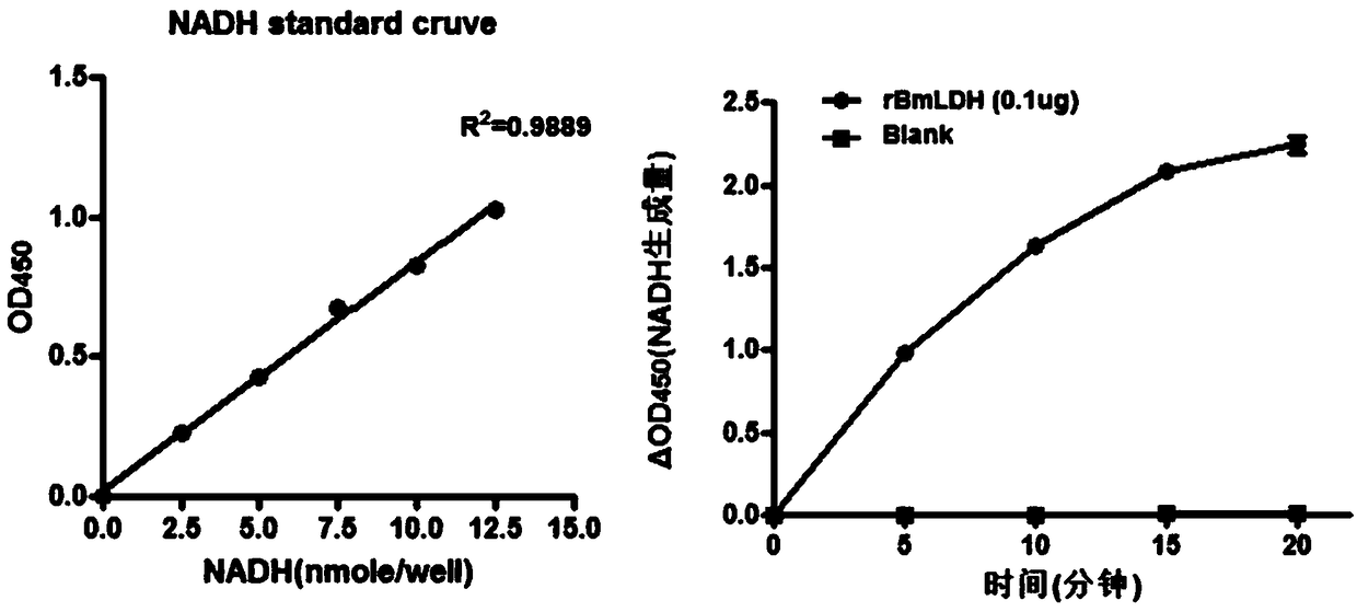
[0048] Embodiment 2: Enzyme activity detection of recombinant rBmLDH protein

[0049] (1) Sample preparation: The rBmLDH protein concentration was determined by the BCA method and the protein concentration was adjusted to 1 mg / mL, and the recombinant protease activity was measured using a protein concentration of 0.1 μg.

[0050] (2) Preparation of LDH standard curve: add 0, 2, 4, 6, 8 and 10uL of 1.25mM NADH standard (in duplicate) into 96-well plate to generate 0 (blank), 2.5, 5, 7.5, 10 and 12.5 nmole / well standard. Add LDH assay buffer to a final volume of 50 µL.

[0051] (3) Experimental procedure for enzyme activity determination: take 48 μL of assay buffer and 2 μL of substrate mixture, and mix well.

[0052] (4) Add 50 μL of enzyme activity reaction mixture to each well, and use horizontal vibration or a pipette to mix well. Avoid light during the experiment.

[0053] (5) After 2-3 minutes, take the initial measurement. Absorbance at 450 nm was initially measured ...

Embodiment 3

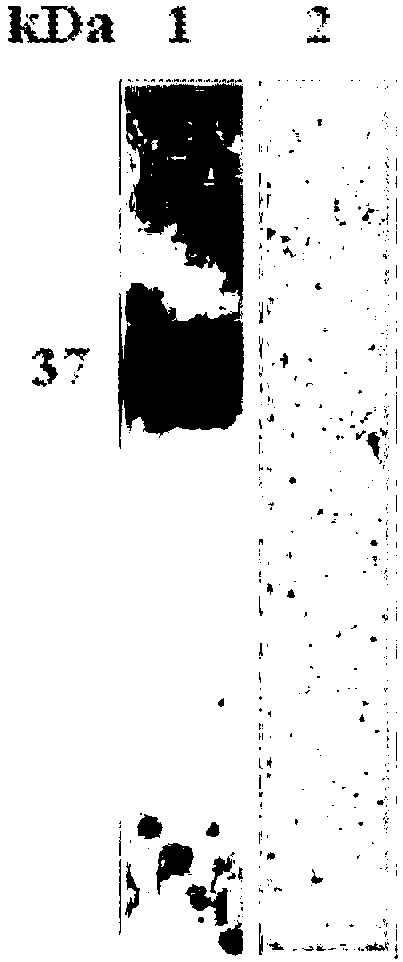
[0055] Example 3: Identification of reactogenicity of recombinant protein rBmLDH

[0056] The purified recombinant protein rBmLDH was first subjected to SDS-PAGE electrophoresis, and then transferred to the NC membrane at a voltage of 50V. After 3h, the NC membrane was blocked with TBST-5% skimmed milk powder for 1h, and washed 3 times with TBST, 5min each time; Put the NC membrane into 1:100 dilution of Babesia microti positive serum and negative serum, incubate at room temperature for 1 hour, wash 3 times with TBST, each time for 5 minutes; add secondary antibody (1:5000 dilution of rabbit anti-mouse IgG), room temperature Incubate for 1 hour, wash 3 times with TBST, and add BCL to develop color after 5 minutes each time. The results of Western blot showed that the purified rBmLDH had a specific reaction at 30kDa with positive sera, but had no reaction with negative sera (see figure 2 ).


PUM


Abstract

The invention discloses a monoclonal antibody resistant to vole babesia. The monoclonal antibody is secreted by a hybridoma cell strain HA5-E4 having a collection number of CCTCC NO: 201835. Aiming atlactic dehydrogenase LDH in a glucide metabolic pathway of the vole babesia, the protein has immunogenicity, and a monoclonal antibody aiming at rBmLDH is prepared; Western blot detection proves thatthe monoclonal antibody is capable of specifically recognizing the vole babesia but cannot recognize the LDH of the host. Therefore, the monoclonal antibody can be applied to detection of vole babesia in infection cells through an immumofluorescence method.

Description

technical field [0001] The invention belongs to the technical field of blood protozoa prevention and treatment of parasitic diseases, and in particular relates to a monoclonal antibody against Babesia microti and its application. Background technique [0002] Babesia microti (Babesia microti) is a kind of obligate parasitic blood protozoan in the erythrocytes of the genus Acroorgan, Sporozoa, Piroplasma, Babesco, and Babesia, and is the cause of Babesia microti in humans and rodents. One of the most important pathogens of entomorrhoids, it is mainly transmitted through the bite of hard ticks, and can also be infected and transmitted through blood transfusion or blood products. Humans or animals show severe clinical symptoms after infection, which can lead to death in severe cases. At present, the disease is widely prevalent in the world, and the cases of human infection are mainly concentrated in the Americas. In recent years, the number of cases has increased sharply, and ...

Claims


Application Information

Patent Timeline
IPC IPC(8): C07K16/40C12N5/20G01N33/577G01N33/569
CPCC07K16/40G01N33/56905G01N33/577G01N2333/904
Inventor 贺兰赵俊龙喻龙战雪燕刘琴赵阳楠周艳琴方瑞申邦胡敏
Owner HUAZHONG AGRI UNIV
Why Eureka
Try Eureka