

Dead/living cell distinguishing fluorescence probe and synthesis method and application thereof
A technology of fluorescent probes and synthesis methods, applied in the direction of fluorescence/phosphorescence, chemical instruments and methods, analytical materials, etc., can solve the problems of lack of distinguishing detection, etc., and achieve the effect of high yield and simple synthesis method
- Summary
- Abstract
- Description
- Claims
- Application Information
AI Technical Summary
Problems solved by technology
Method used
Image

Examples
Embodiment 1
[0032] Example 1 Synthesis of fluorescent probes.
[0033] (1) Synthesis of 1,2-dimethyl-quinoline iodide salt (compound 1):
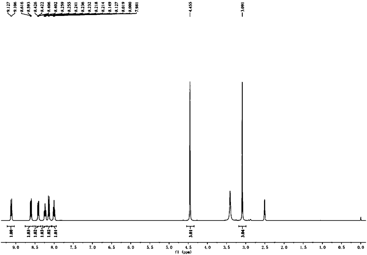
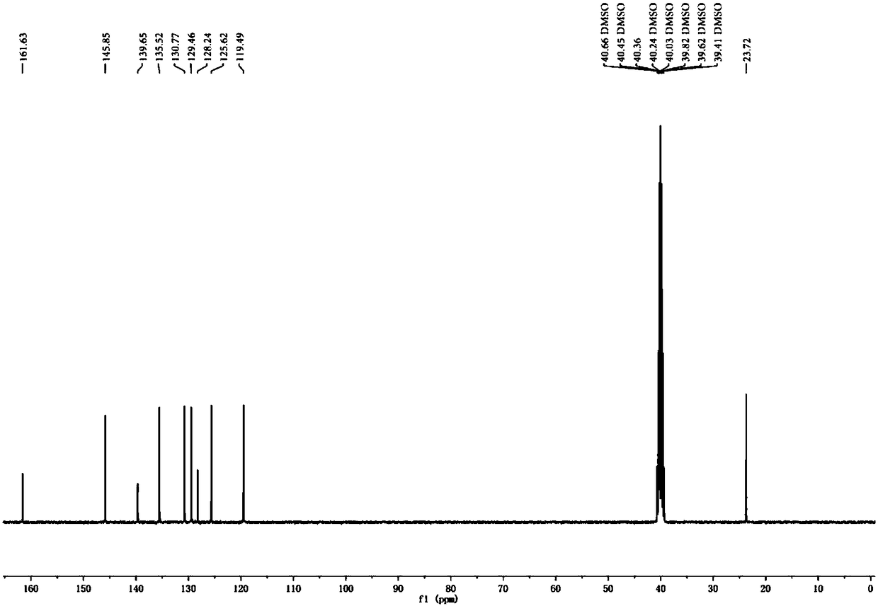
[0034] Add 5-10 mL of ethanol to the round bottom flask, then add 1.1-1.4 mL of 2-methylquinoline, add 0.5-1.2 mL of iodomethane solution, heat to 40-60 degrees for 24-48 hours, and the reaction is complete After cooling the reaction system to room temperature, a solid precipitated out, filtered and washed with ethanol to obtain 1,2-dimethyl-quinoline iodide salt (compound 1), with a yield of 92%. Its H NMR spectrum is as figure 1 shown. 1 H NMR (400 MHz, DMSO- d 6 ) δ 9.12 (d, J = 8.5 Hz, 1H), 8.60 (d, J = 9.0 Hz, 1H), 8.41 (dd, J = 8.2, 1.6 Hz, 1H), 8.24 (ddd, J = 8.8, 7.0, 1.6Hz, 1H), 8.14 (d, J = 8.5 Hz, 1H), 8.00 (t, J = 7.6 Hz, 1H), 4.45 (s, 3H),3.09 (s, 3H). 13 C NMR (101 MHz, DMSO- d 6 ) δ (ppm): 161.63, 145.85, 139.65, 135.52, 130.77, 129.46, 128.24, 125.62, 119.49, 40.66, 40.45, 40.36, 40.24, 40.03, 2, 39.63, 39.62
[0035...
Embodiment 2
[0037] Example 2 Cell imaging of fluorescent probe MNQI.
[0038](1) Cell culture, treatment and staining:
[0039] Set the density to 3 x 10 5 cells / mL of HeLa cells were inoculated into sterile 35 mm imaging culture dishes in CO 2 Incubator (at 37°C, 5% CO 2 ) for more than 12 hours to allow the cells to adhere to the wall. Adherent cells were treated with 4% paraformaldehyde for 30 min to obtain dead cell samples. The DMSO solution of the probe obtained in Example 1 was prepared at a concentration of 2.5 mM as the mother solution, and the mother solution was added to the dead and living cell culture dish so that the final concentration was 5 μM. Continue to culture under the same conditions for 1 h, then aspirate the cell culture medium, wash the cells with the medium for 3 times, and then perform the cell imaging experiment.
[0040] (2) Confocal microscope imaging:
[0041] With 488 nm as the excitation wavelength, the collection wavelength of the green light chann...
Embodiment 3
[0042] Example 3 Probe MNQI co-localizes with commercial probes.
[0043] In order to further confirm the coloring position of the probe MNQI in dead and living cells, the commercial mitochondrial dye Mitochondrial Deep Red (MTDR) and the commercial nuclear dye (Hoechst33342) were used to perform colocalization staining imaging with MNQI in living cells and dead cells, respectively.
[0044] In the cell colocalization experiment, the cells were first stained with 200 nM MTDR for 30 min, and then 5 μM MNQI was added to stain the cells for 60 min, then the cell culture medium was aspirated, and the cells were washed with the medium for 3 times for cell imaging. Use 488 nm as the excitation wavelength, collect the fluorescence at 570-620 nm to collect the fluorescence signal of MNQI; use 633 nm as the excitation wavelength, collect the fluorescence at 663-738 nm to collect the fluorescence signal of MTDR. Get the fluorescence picture as Figure 6 The first column shows, from lef...
PUM
 Abstract
Description
Claims
Application Information